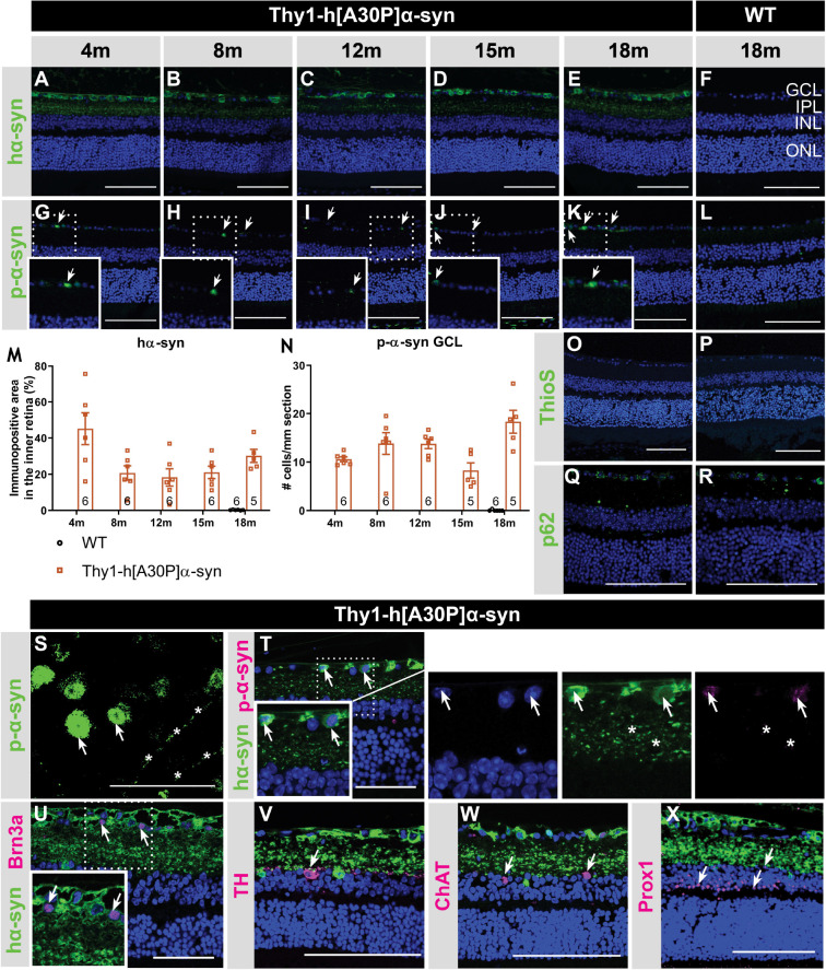
FIGURE 1.
Inner retinal hα-syn expression is accompanied by α-syn phosphorylation, yet no ThioS positive aggregation or p62 accumulation, in the retina of Thy1-h[A30P]α-syn mice. Representative images of hα-syn immunostainings (A–E); p-α-syn immunostainings (G–K); and ThioS staining (O) on retinal sections of α-syn mice at 4, 8, 12, 15 and 18 months of age. (F,L,P) No staining was observed in the WT controls, at any age (only 18 months shown here). (M,N) Quantitative analysis of the hα-syn fluorescent area and counting of the p-α-syn positive cells did not reveal an increase of hα-syn expression in the inner retina or p-α-syn cell density in α-syn mice with age. (O,P) No ThioS positive inclusions were found in the retina of transgenic nor wild type animals in any of the age groups. (Q,R) No difference in retinal p62 accumulation or localization was detected between transgenic and wild type animals at 18 months of age. (S) p-α-syn immunostaining on a retinal wholemount of an α-syn mouse showed p-α-syn localization in cell bodies (arrows) and neurites (asterisks). (T) Double staining of hα-syn with p-α-syn revealed clear colocalization. (U–X) Double staining of hα-syn with Brn3a, TH, ChAT and Prox1 revealed expression of Brn3a in hα-syn positive cells, yet no colocalization in dopaminergic and cholinergic cells. Scale bar: 100 μm (A–R, V–X) or 50 μm (S–U); GCL, ganglion cell layer; INL, inner nuclear layer; IPL, inner plexiform layer; and ONL, outer nuclear layer.

