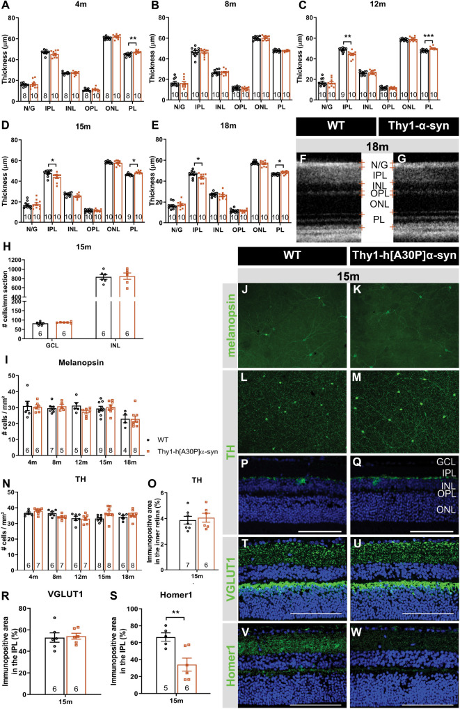
FIGURE 2.
Outer retinal thickening and inner retinal thinning, associated with loss of postsynaptic labeling, in Thy1-h[A30P]α-syn mice. (A–E) Longitudinal OCT measurements in 4- (A), 8- (B), 12- (C), 15- (D), and 18-month-old (E–G) mice, revealed significant differences in retinal layer thickness between α-syn and WT mice of 4 months (PL thickening), 15 months (PL thickening and IPL thinning), and 12 and 18 months of age (PL thickening and IPL thinning). (H) Cell counts on hematoxylin and eosin-stained sections in the GCL and in the INL did not reveal significant differences between transgenic animals and WT controls at 15 months of age. (I–W) Representative images of retinal wholemounts stained for melanopsin (J,K) and TH (L,M), and of retinal sections stained for TH (P,Q), VGLUT1 (T,U), and Homer-1 (V,W), of 15-month-old α-syn and WT mice. Counting the number of melanopsin- (I) and TH- (N) positive cells on retinal wholemounts revealed no significant differences between transgenic and WT animals. No significant differences were uncovered in TH plexus (O) and VGLUT1 (R) immunopositive area, yet a strong decrease of the Homer1 (S) signal was seen. Scale bar: 100 μm; Two-Way ANOVA with Tukey multiple comparisons post hoc test (I–N). Unpaired t-test (per retinal layer; A–F,O,R,S): *p < 0.05; **p < 0.01; and ***p < 0.001. N/G, retinal nerve fiber layer + GCL; GCL, ganglion cell layer; INL, inner nuclear layer; IPL, inner plexiform layer; ONL, outer nuclear layer; OPL, outer plexiform layer; and PL, photoreceptor layer.

