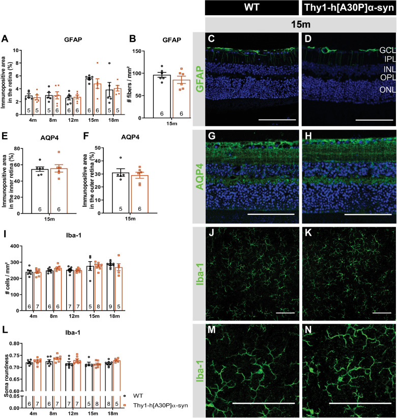
FIGURE 4.
Macroglia and microglia reactivity and water homeostasis appear normal in Thy1-h[A30P]α-syn mice. Representative images of retinal cross-sections stained for GFAP (C,D) and wholemounts stained for Iba-1 (J,K,M,N) and cross-sections stained for AQP4 (G,H) in 15-month-old α-syn and WT mice. (A,B) When measuring the GFAP immunopositive area and the number of radial fibers in the inner retina, no differences in macroglia reactivity were uncovered between transgenic and WT animals in any of the age groups. (I,L) No differences in Iba-1+ cell density and cell soma roundness, indicative of microgliosis, were observed. (E,F) AQP4 immunopositive area or localization in the inner versus outer retina of α-syn mice versus age-matched WT animals was similar. Two-Way ANOVA with Sidak’s multiple comparisons post hoc test (A,I,L) or unpaired t-test (B,E,F). Scale bar: 100 μm.

