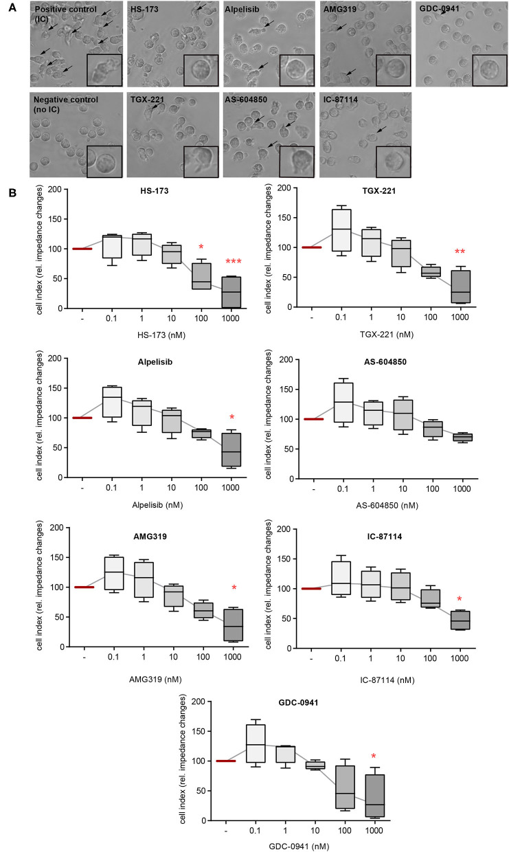
Figure 2.
IC-induced spreading of PMNs mainly depends on PI3Kδ. Freshly isolated human blood PMNs were activated with immobilized ICs in the presence of either one of the PI3K isoform-selective inhibitors. (A) Microscopic appearance of PMNs 2 h after IC-stimulation. IC-stimulated PMNs show clear spreading on surface (arrows). The highest concentration of all inhibitors (1 μM) reduced the adhesion of PMNs. (B) Spreading was monitored live for 2 h and the area under curve (AUC) was analyzed. Data are shown as Tukey's box-and-whisker plots. n = 4. ANOVA on ranks (Kruskal-Wallis) was applied followed by a Bonferroni t-test for multiple comparisons. Data were normalized to IC-stimulated cells. *p < 0.05, **p < 0.01, ***p < 0.001.

