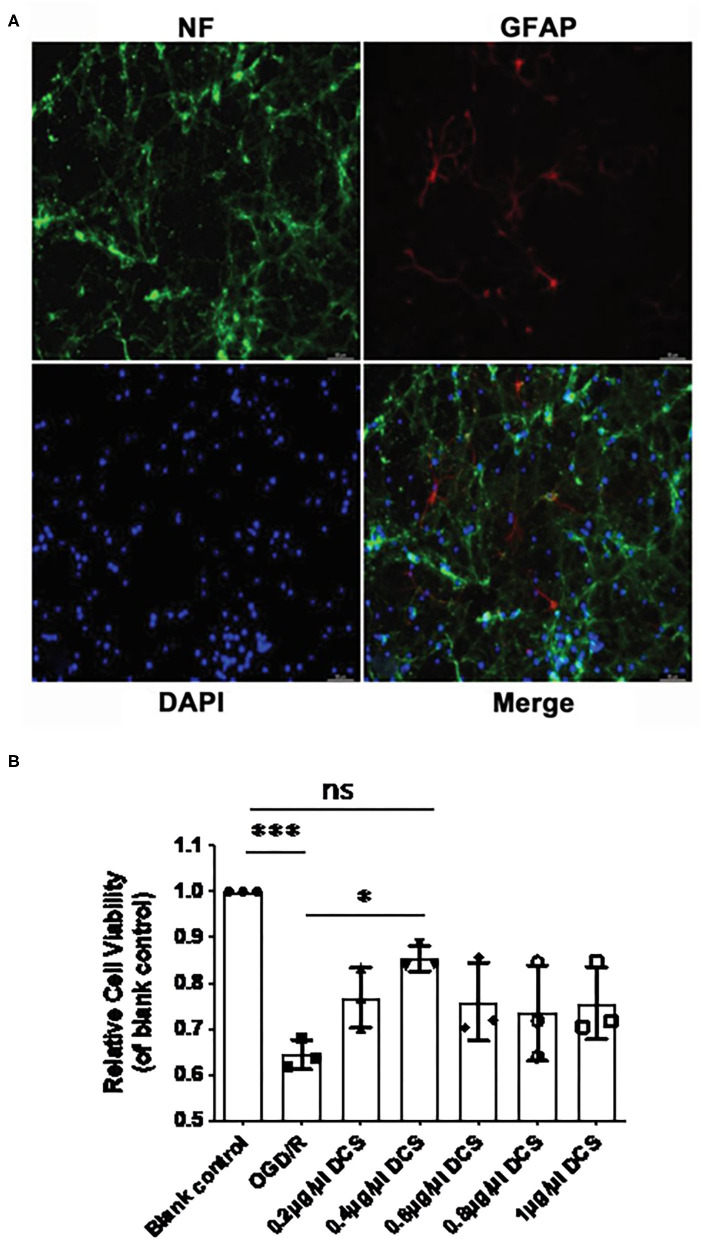
Figure 2.
Effect of DCS on the viability of cultured primary neurons exposed to oxygen-glucose deprivation/reperfusion (OGD/R). (A) Cultured primary neurons isolated from rat E18 embryos and immunostained for neurofilament (NF, expressed in neurons; green) and glial fibrillary acidic protein (GFAP, expressed in astrocytes; red). Nuclei were stained with 4′,6-diamidino-2-phenylindole (DAPI; blue). Neuronal cell purity exceeded 95%. (B) Cell viability determined by using Cell Counting Kit-8. Primary neurons were exposed to OGD/R and administered DCS for 4 h at various concentrations (0, 0.2, 0.4, 0.6, 0.8, and 1 μg/μl). Cells not exposed to OGD/R were used as a blank control group. Data shown as mean ± SEM, n = 3. *p < 0.05, ***p < 0.0005.

