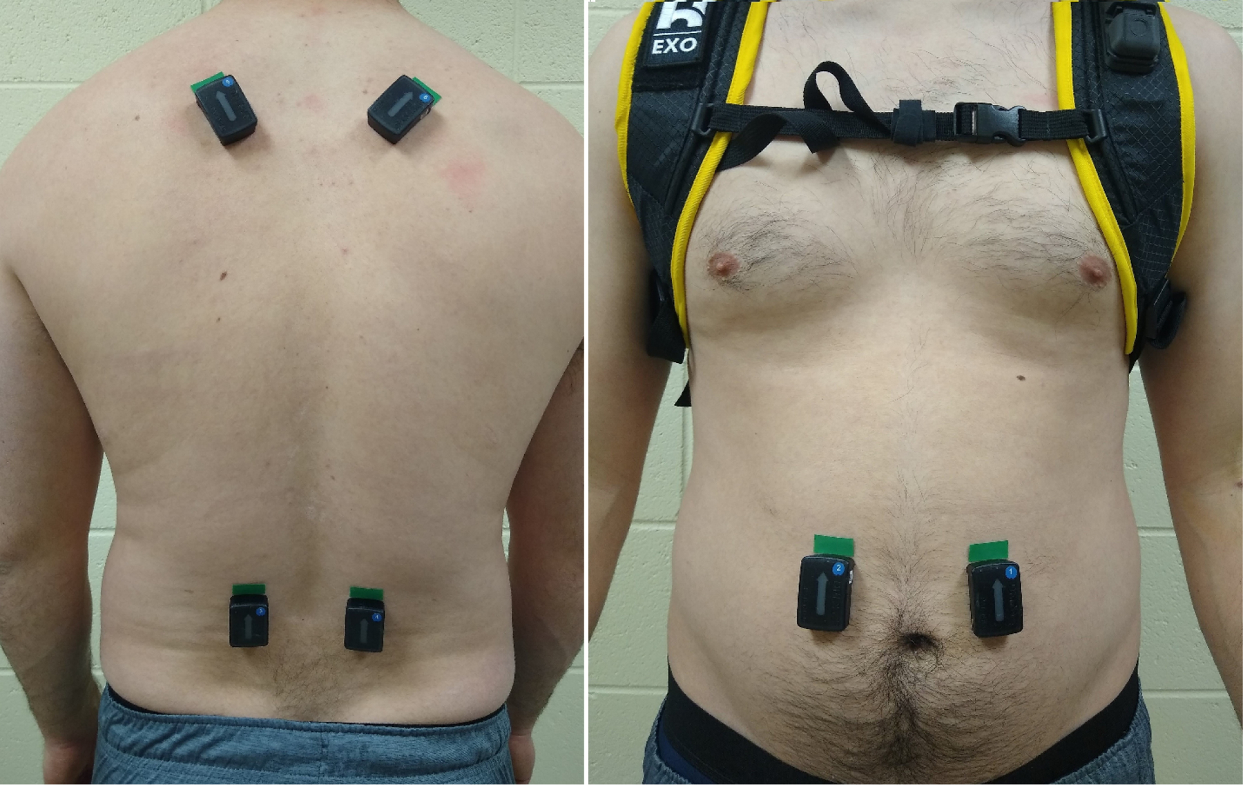
Figure 3.

Close-up of the electromyography sensors on the back (bottom: erector spinae; top: middle trapezius) and front (rectus abdominis). Prior to beginning the study protocol, additional tape was placed over the sensors for better fixation, and a shirt and the full exosuit were donned.
