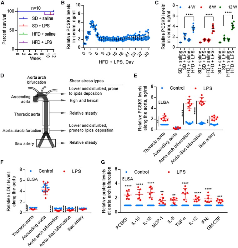
Figure 4.
Distribution of PCSK9 expression along the aorta. (A) Rabbit survival rate in different groups with or without LPS treatment. (B) In HFD groups, PCSK9 levels in first 30 days after LPS treatment. Data were normalized to HFD + saline, set control HFD + LPS = 1 (0 day). (C) LPS treatment significantly induces serum PCSK9 levels in both SD and HFD groups, measured by ELISA at 4, 8, and 12 weeks. (D) Schematic drawing of flow patterns along the aorta. (E) PCSK9 levels along the aorta with or without LPS treatment on day 3 HFD, measured by ELISA. LPS induces PCSK9 expression in thoracic aorta, aorta arch branch points, aorta–iliac bifurcation, and iliac artery but not in ascending aorta. PCSK9 levels are highest in aortin arch branch points and aorta–iliac bifurcation. (F) ELISA analysis of LDLr expression along the aorta with or without LPS treatment on day 3 HFD. (G) ELISA analysis for expression of PCSK9 and pro-inflammatory cytokines at aorta arch bifurcation on day 3 HFD, set control without LPS = 1 in each group. Bar graphs represent data compiled from three independent experiments (n = 7 rabbit per genotype), shown as mean ± standard deviation. The significances between two groups were tested by unpaired t-test; Multiple comparisons were analysed by one-way ANOVA, followed by Tukey’s post hoc comparisons test (**P < 0.01; ****P < 0.0001).

