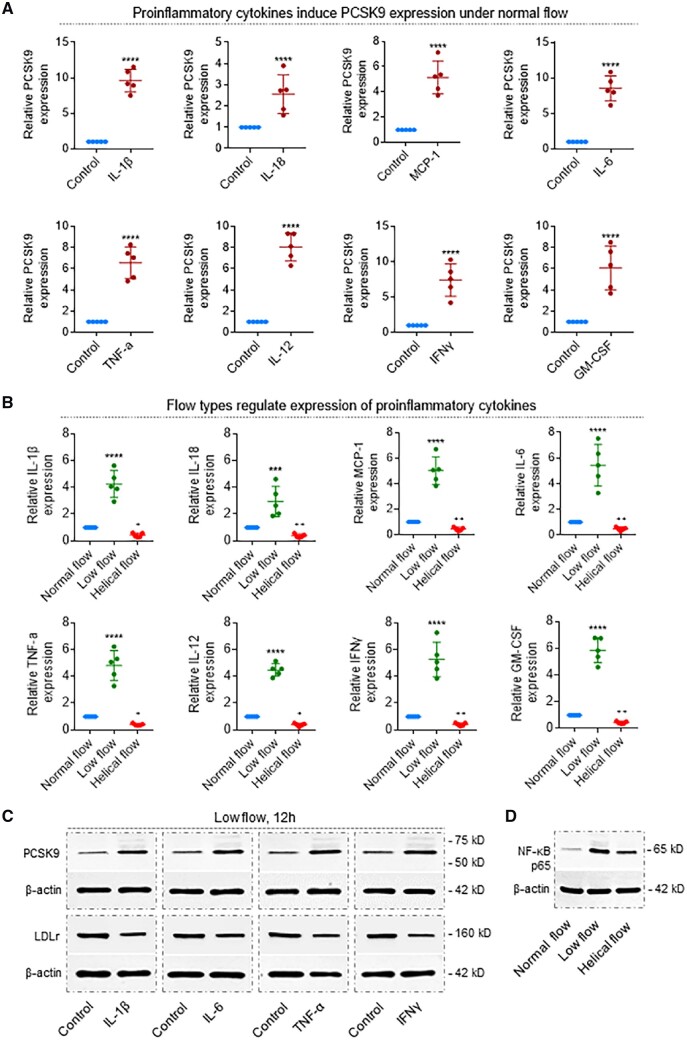
Figure 6.
Pro-inflammatory cytokines determine PCSK9 levels, which is regulated by flow patterns. (A) Recombinant proteins IL-1β, IL-18, MCP-1, IL-6, TNFα, IL-12, IFNγ, and GM-CSF induce PCSK9 expression in the rabbit thoracic aorta. Recombinant proteins were added to the perfusion medium in different concentrations. Aortas were perfused for 12 h under normal flow conditions. ****P < 0.0001. Bar graphs represent data compiled from three independent experiments (n = 5 rabbit per genotype), shown as mean ± standard deviation. (B) Helical flow inhibits whereas low flow enhances LPS-induced release of pro-inflammatory cytokines: IL-1β, IL-18, MCP-1, IL-6, TNFα, IL-12, IFNγ, and GM-CSF. ****P < 0.0001 vs. normal group; + P < 0.05, + P < 0.05 vs. normal group, ++ P < 0.01 vs. normal group. (C) Pro-inflammatory cytokines (IL-1β, IL-6, TNF-α, and IFNγ) regulate expression of PCSK9 and LDLr in the rabbit thoracic aorta. Aortas were perfused for 12 h under low-flow conditions, treatment with IL-1β at 4 ng/mL, IL-6 at 20 ng/mL, TNF-α at 50 ng/mL, and IFNγ at 10 ng/mL. (D) Flow types regulate NF-κB expression in the rabbit thoracic aorta. Aortas were perfused under low-flow conditions, treatment with LPS at 20 ng/mL in the perfusion medium for 12 h. Bar graphs represent data compiled from three independent experiments (n = 5 rabbit per genotype), shown as mean ± standard deviation. The significances between two groups were tested by unpaired t-test; Multiple comparisons were performed by one-way ANOVA, followed by Tukey’s post hoc comparisons test.

