Figure 2.
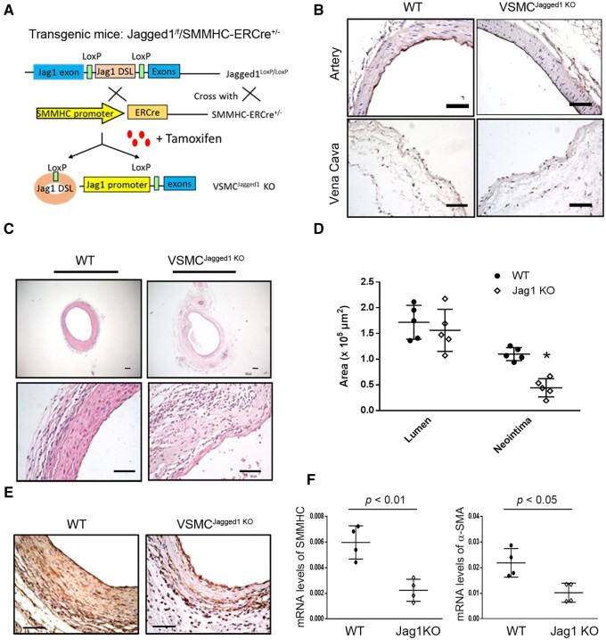
Jagged1 KO in VSMCs suppresses vascular remodelling in AVGs. (A) The schematic for generating VSMC-specific Jagged1 KO mice. (B) Jagged1 expression was determined in control vein and arteries from WT and VSMCJagged1 KO mice by immunostaining. (C and D) Representative images of H&E staining (C) and the areas of the lumen and neointima from of the 1 month AVGs created in VSMCJagged1+/+ and VSMCJagged1 KO mice (D) (n = 5; two sample t-test was used for statistical analysis; scale bars are 50 µm in all panels). (E) Representative images of the immunostaining of α-SMA in AVGs from WT and VSMCJagged1 KO mice. (F) The expression of VSMC markers were determined by RT–PCR in AVGs from WT and VSMCJagged1 KO mice. The Ct values for SMMHC in AVGs created in WT and Jagged1 KO mice are 29.7 ± 1.52 and 31.1 ± 2.6). The Ct values for α‐SMA in AVGs created in WT and Jagged1 KO mice are 27.91 ± 2.38 and 28.6 ± 2.65) (n = 4, *, P < 0.05 compared with results of WT; t-test was used for statistical analysis).

