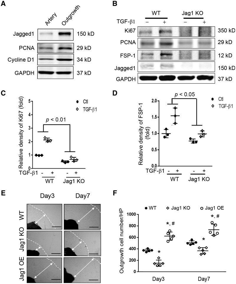
Figure 3.
Expression of Jagged1 promoted SMC phenotype switch from a quiescent to the synthetic status: (A) Common carotid arteries were dissected, half of the arteries were cultured, and half were kept for later use. After 7 days, proteins from the artery and outgrowth SMCs were isolated and the western blot was performed (n = 3), depicting gain of Jagged1 expression and increasing expression of PCNA and Cyclin D1 in the outgrowth SMCs. (B) VSMCs from WT and VSMCJagged1 KO mice were treated with or without 2 ng/ml TGF-β1 for 24 hrs. The expressions of Ki67, PCNA, and FSP-1 were detected by western blot. (C and D) Quantitation analysing the densities of Ki67 and FSP-1. Representative data from three experiments. Two-way ANOVA with an interaction between treatment and Jagged1 expression levels was used to test the expression of Ki67 and FSP-1 changes among the two groups. The effects of TGF-β1 on the expression of Ki67 and FSP1 were different in Jagged1 KO VSMCs compared to WT cells treated with TGF-β1. (E) Artery segments from WT, Jagged1 KO mice, and Jagged1 OE mice were seeded onto 24-well plates ex vivo and photographs were taken at indicated time points. Representative pictures are presented. The dashed lines indicate the edge of the cells, and the double arrowed lines show the distance of outgrowth cells (scale bars are 200 µm). (F) The cell numbers in panel (E) were counted and summarized (*, P < 0.05 vs. WT; #, P < 0.05 vs. Jagged1 KO; one-way ANOVA was used for statistical analysis for day 3 or day 7 separately, n = 5).

