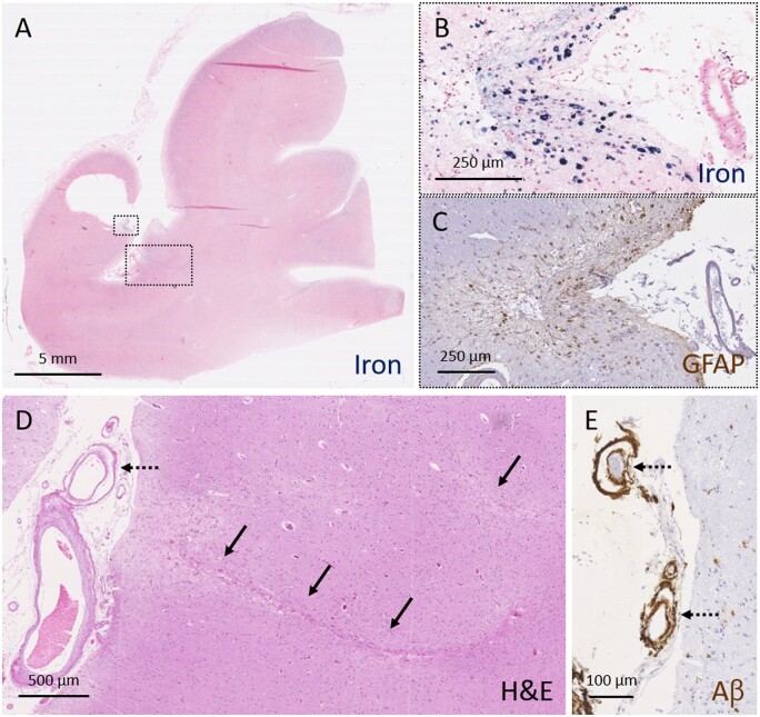
Figure 3.
Moderate-to-severe cSS is associated with advanced leptomeningeal CAA and secondary tissue injury. A representative example of severe cSS as assessed on a Perls’ Prussian blue-stained section (A). Higher magnification of the area outlined with the small box in A, reveals greater detail of severe cSS (B). An adjacent section stained for GFAP revealed many reactive astrocytes in the superficial cortical layers in this area (C). Two cortical microinfarcts (indicated with solid arrows) were observed in the neighbouring cortical area (D, area corresponds to larger box in A). Note the presence of vessel-within-vessel pathology (indicated with broken arrows) in combination with severe CAA of the leptomeningeal vessels (D and E). Aβ = amyloid-β; H&E = haematoxylin and eosin.

