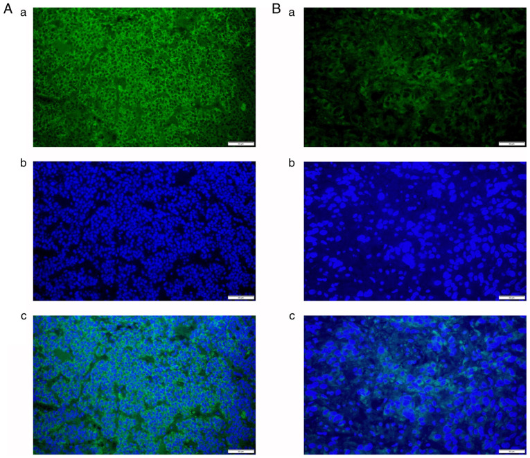
Figure 1.
(A) Representative immunofluorescence signals of (A-a) COLI and (A-b) DAPI in firm tumors; (A-c) merged image of A-a and A-b. (B) Representative immunofluorescence signals of (B-a) COLI and (B-b) DAPI in soft tumors; (B-c) merged image of B-a and B-b (magnification, x400; scale bars, 50 µm). COLI, collagen type I.

