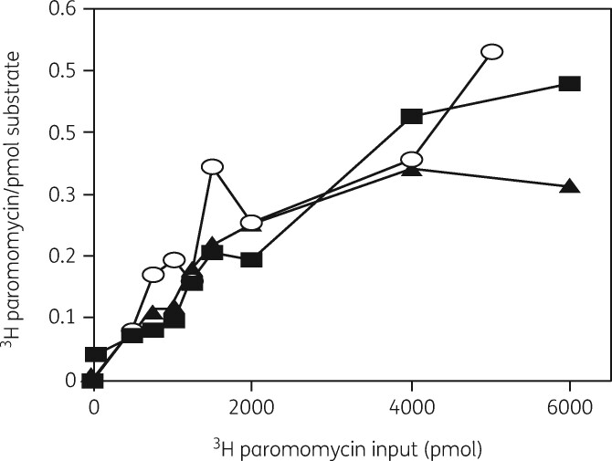
Figure 12.
Binding of [3H]-paromomycin to 30S and 50S subunits and the 21S precursor particle. A filter-binding assay with increasing amounts of [3H]-paromomycin was used to measure the binding interaction. [3H]-paromomycin binding to 30S subunits (filled squares), 50S subunits (filled triangles) and to the 21S precursor (open circles). Reproduced with permission from Springer Nature, reference 136.

