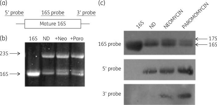
Figure 13.
Identification of precursor forms of 16S rRNA in antibiotic-treated cells. (a) Location of the hybridization probes used. (b) Gel pattern of rRNA from control and drug-treated cells. (c) Northern blot of rRNA from antibiotic-treated cells. Top: hybridization with internal 16S rRNA-specific probe. Middle: hybridization with a 5′ precursor sequence-specific probe. Bottom: hybridization with a 3′ precursor sequence-specific probe. ND, control. Reproduced with permission from Springer Nature, reference 136.

