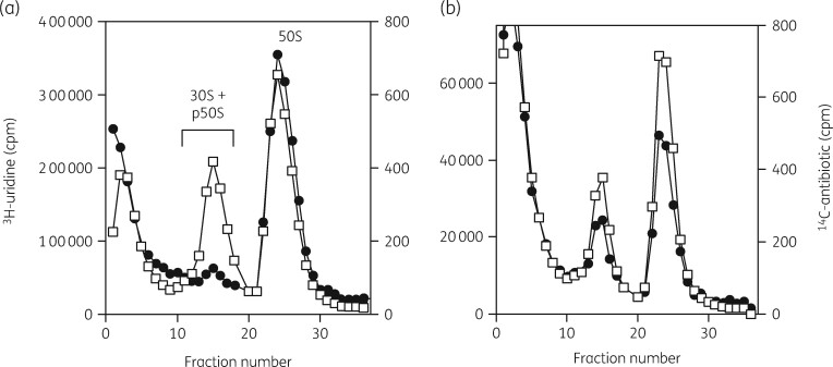
Figure 5.
Sucrose gradient profiles of ribosomal subunits from cells grown with [3H]-uridine- and [14C]-labelled antibiotics. (a) Profile showing [3H]-uridine-labelled subunits (open squares) and [14C]-erythromycin (closed circles) bound to both 50S subunits and to a 32S precursor particle. (b) Profile showing [3H]-uridine-labelled subunits (open squares) and [14C]-azithromycin (closed circles) bound to both 50S subunits and to a 32S precursor particle. Reproduced with permission from Bentham Science Publishers Ltd from reference 92.

