Figure 4.
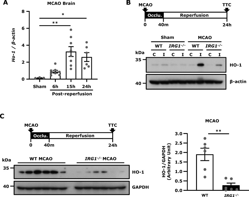
IRG1 deficiency represses HO-1 expression in the ischaemic brain. (A) WT mice were subjected to sham or 40 min MCAO. The ipsilateral hemispheres of sham controls were harvested at 15 h post-reperfusion (n = 3 mice), and the ipsilateral hemispheres of MCAO mice were harvested at 6, 15 and 24 h post-reperfusion (n = 6–8 mice per time point). Samples were then subjected to Q-PCR analysis for HO-1 expression. *P < 0.05, **P < 0.01 by one-way ANOVA. (B) WT and IRG1−/− mice were subjected to sham or 40 min MCAO. At 24 h post-injury, the contralateral and ipsilateral hemisphere tissues were harvested and subjected to western blot analysis for HO-1 expression. The representative results of HO-1 expression in the contralateral (C) and ipsilateral (I) hemispheres of sham and MCAO mice are shown. Similar results were observed in three independent experiments. (C) The ischaemic brains were harvested from WT and IRG1−/− MCAO mice at 24 h post-injury, and the ipsilateral hemisphere tissues were then subjected to western blot analysis for HO-1 expression. The level of HO-1 expression was also quantified (n = 6 mice per group). **P < 0.01 by Mann–Whitney U test.

