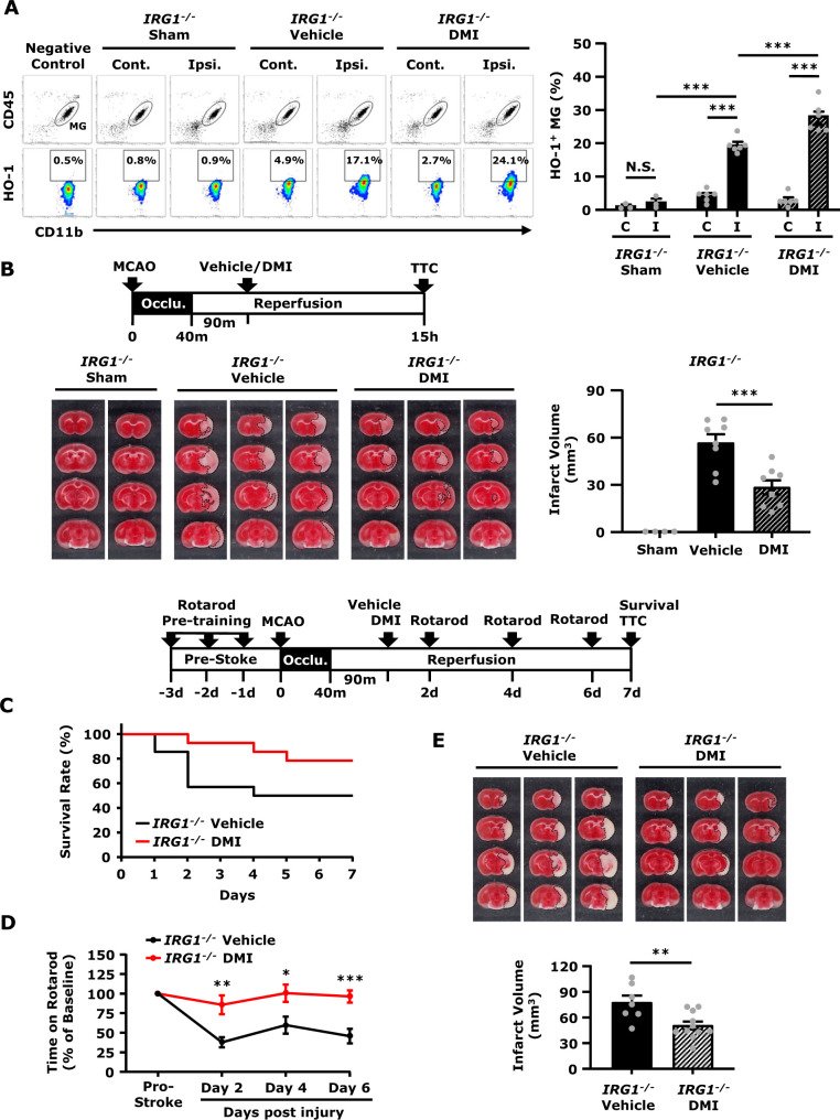
Figure 7.
DMI rescues IRG1 deficiency-exacerbated brain injury in ischaemic stroke.IRG1−/− mice were subjected to sham or 40 min MCAO. IRG1−/− MCAO mice were intraperitoneal administered with vehicle or DMI 500 mg/kg at 90 min post-reperfusion. (A) At 15 h post-injury, the ischaemic brains harvested from sham (n = 3 mice) and vehicle- and DMI-treated IRG1−/− MCAO mice (n = 6 mice per group) were separated into the contralateral (Cont.; C) and ipsilateral (Ipsi.; I) hemispheres followed by mononuclear cell isolation. The isolated cells were then stained with antibodies of CD45 and CD11b followed by intracellular staining of primary HO-1 antibody and secondary Alexa Fluor 546 antibody. The cells were then subjected to flow cytometry analysis. Negative control (secondary antibody only) was used as a negative control to determine CD45intCD11b+ MG positive for HO-1 expression. ***P < 0.001, N.S., no significant differences by two-way ANOVA. (B) At 15 h post-injury, the ischaemic brains harvested from sham (n = 4 mice) and vehicle- and DMI-treated IRG1−/− MCAO mice (n = 8 mice per group) were subjected to TTC staining. The representative TTC-stained samples of sham (n = 2 mice) and vehicle- and DMI-treated MCAO mice (n = 3 mice per group) are shown. The infarct volumes were also measured. ***P < 0.001 by one-way ANOVA. (C–E) IRG1−/− MCAO mice were intraperitoneal administered with vehicle or DMI 500 mg/kg at 90 min post-reperfusion (n = 14 mice per group). Vehicle- and DMI-treated IRG1−/− MCAO mice were subjected to (C) survival assay up to Day 7 post-injury and (D) rotarod tests at Day 2, 4 and 6 post-injury. *P < 0.05, **P < 0.01, ***P < 0.001 by unpaired t-test. (E) At Day 7 post-injury, the survived vehicle- (n = 7 mice) and DMI- (n = 11 mice) treated IRG1−/− MCAO mice were sacrificed and subjected to TTC staining. Three representative TTC-stained samples are shown. The infarct volumes were also measured. **P < 0.01 by unpaired t-test.

