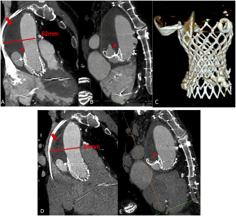
Figure 5.
(A–C) Multi-slice computed tomography 4 weeks after endovascular entry closure. Arrow indicates the false lumen now filled with thrombus. Arrowhead indicates a small residual perfusion. The aortic diameter is unchanged. (C) Three-dimensional reconstruction showing the Occluder fixed at the supra-annular nitinol frame of the Evolut valve. (D and E) Multi-slice computed tomography 12 weeks after endovascular closure. Arrow indicates the false lumen filled with thrombus. Arrowhead indicates the residual perfused lumen now reduced by half. The diameter of the aorta is reduced by 7–55 mm due to thrombus shrinkage.

