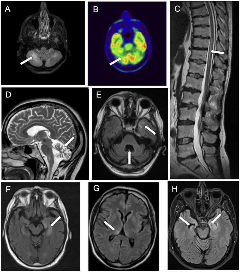
Figure 3.
Neuroimaging findings in patients with KLHL11-Abs. Axial brain MRI (A), showing hypersignal asymmetrically involving both cerebellar hemispheres, colocalizing with an area of cerebellar hypometabolism on brain PET (B). Sagittal spine MRI showing spinal cord hypersignal in a patient with myelitis (C). Marked cerebellar atrophy is shown in both sagittal (D) and axial (E) brain MRI. The atrophy is more pronounced in the vermis and associates with an area of hypersignal involving the temporal lobe (E). Axial brain MRIs showing hypersignal involving the left mesial temporal lobe (F), the right thalamus (G) and coexistence of left mesial temporal lobe hypersignal and right hippocampal atrophy.

