Figure 5.

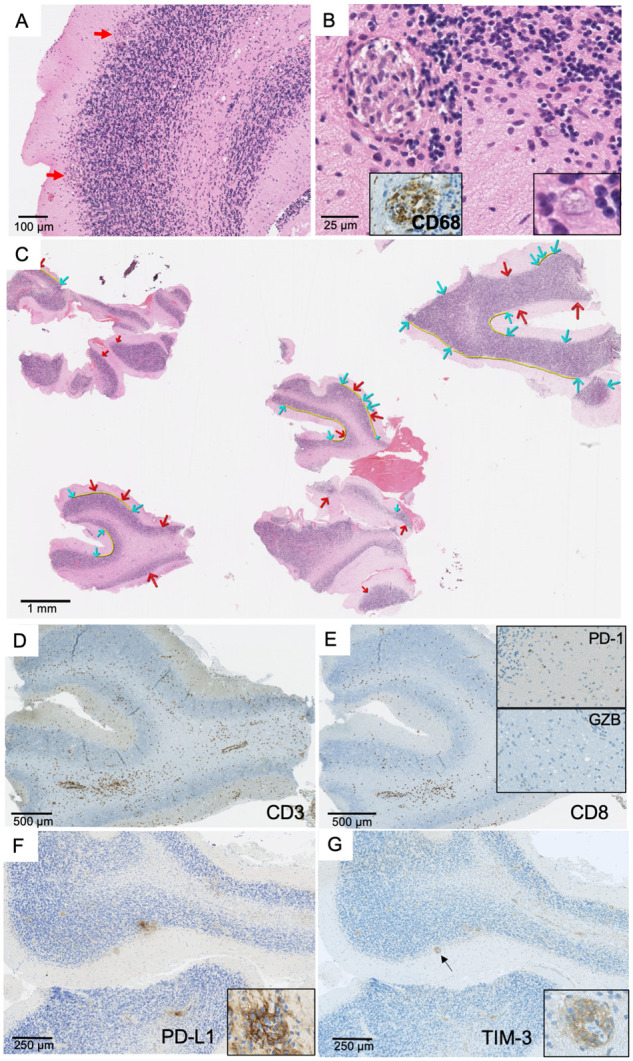
Brain biopsy in a patient with KLHL11-Ab encephalitis. Pathological brain findings demonstrating the presence of granulomas located at the interface between granular and molecular layers of the cerebellum (A, ×100). These granulomas were formed by epithelioid cells showing positive CD68 staining (B, left panel, ×200) in proximity to Purkinje cells with shrunk cytoplasm (B, right panel, ×300 and ×500). Panel C (×10): a huge neuronal loss was demonstrated by the increased distance (yellow line) between Purkinje cells (blue arrows); numerous granulomas were also observed (red arrows). CD3+ T cells infiltrate was observed predominantly in perivascular space (D, ×50) with numerous CD8+ T cells (E, ×50). Scattered lymphocytes expressing PD-1 (E, top insert, ×100) but not Granzyme B (E, bottom insert, ×150) were detected. A strong expression of PD-L1 was observed in granulomas (F, ×100), which was formed by macrophages (F, insert, ×400). A strong expression of TIM-3 was also detected in granulomas (arrow) (G, ×100; insert: ×400).
