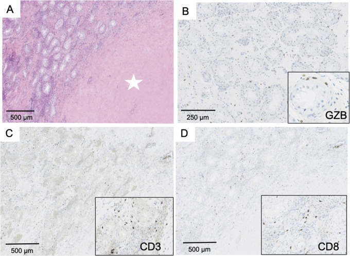
Figure 6.
Pathological findings in ‘burned-out’ testicular tumours. Panel A (×100) shows a large area of fibrosis with no tumour cells (white star), surrounded by testicular parenchyma with abundant lymphocytic infiltrate. Numerous T cells expressed Granzyme B (B, ×200; insert: ×300). The infiltrate was represented by abundant T CD3+ cells (C, ×100, insert: ×200), most commonly by CD8+ (D, ×100; insert: ×200).

