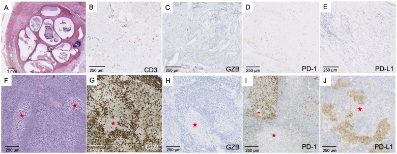
Figure 7.
Testicular tumour and lymph node metastasis. Testicular tumour (A–E). Partial fibrous involution of a testicular tumour resembling a teratoma (A, ×10) with staining of CD3+ T cells (B, ×150), Granzyme B+ cells (C, ×150), few scattered PD-1+ cells (D, ×100) and no expression of PD-L1 within the teratoma (E, ×150). Lymph node metastasis (F–J). Metastasis corresponding to the localization of a seminoma (red star: tumour) (F, ×100), with abundant CD3+ T cell infiltrate surrounding tumour cells (G, ×100), no Granzyme B expression (H, ×100), strong PD-1 expression by lymphocytes (I, ×100) and strong PD-L1 expression preferentially observed in macrophages (J, ×100).

