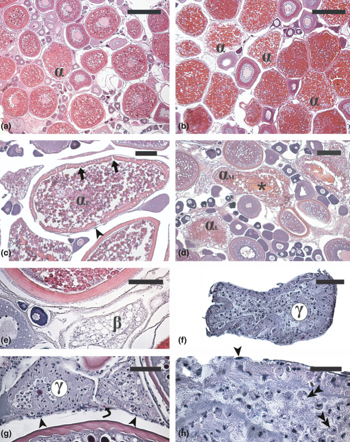
FIGURE 1.

Micrographs of ovary sections from adult Atlantic bluefin tuna Thunnus thynnus (a, b, f, g and h), swordfish Xiphias gladius (e) and greater amberjack Seriola dumerili (c and d) in different phases of the reproductive cycle. (a) Advanced vitellogenic ovary showing a physiological rate of atresia. (b) Extensive atresia of vitellogenic follicles in a specimen that underwent an acute stressing event described in Corriero et al. (2011). (c) Early α‐atretic vitellogenic follicle (αE) characterized by zona radiata fragmentation and nucleus disappearance. (d) Mid (αM) and late (αL) atresia of vitellogenic follicles characterized by progressive zona radiata digestion and yolk granule coalescence. (e) β‐atretic follicle characterized by numerous lipid vesicles and total reabsorption of yolk granules. (f) Early and (g) late γ atretic follicles showing a progressive reduction of the number of follicular cells. (h) Particular of a late γ‐atretic follicle showing follicular cells in active phagocytosis. Haematoxylin‐eosin staining. Magnification bars: 400 µm in (a) and (b); 100 µm in (c) and (e); 150 µm in (d); 50 µm in (f) and (g); 30 µm in (h). α, α‐atretic vitellogenic follicle; β, β‐atretic follicle; γ, γ‐atretic follicle; arrow, zona radiata breakdown; arrowhead, thecal cell; double arrowhead, follicular cell in active phagocytosis; asterisk, residual zona radiata under digestion; curved arrow, blood vessel
