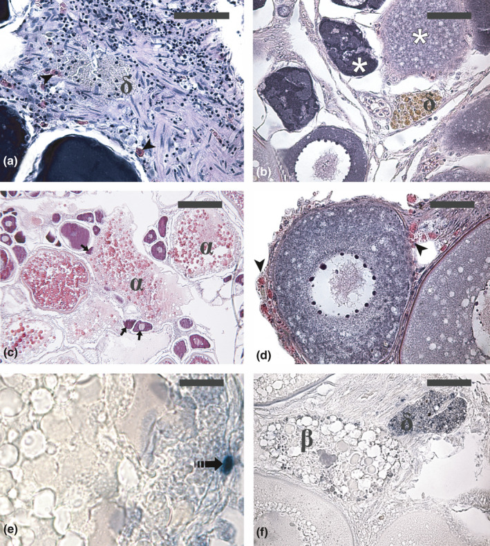
FIGURE 2.

Micrographs of ovary sections from adult Atlantic bluefin tuna Thunnus thynnus (a, c, e and f) and swordfish Xiphias gladius (b and d) in different phases of the reproductive cycle. (a) δ‐atretic follicle showing yellow‐pigmented granules. (b) δ‐atretic follicle showing cells containing brownish pigments. (c) Degenerating unyolked follicles incorporated by an atretic vitellogenic follicle (arrow). (d) Eosinophilic granulocytes at the periphery of an early atretic previtellogenic follicle (arrowhead). (e) Apoptotic granulosa cell in an early α‐atretic vitellogenic follicle (dashed arrow). (f) Apoptotic cells and bodies (dark dots) in β and δ atretic follicles. Haematoxylin‐eosin staining in (a–d). Staining of apoptotic cells and bodies by the terminal deoxynucleotidyl transferase‐mediated 2′‐deoxyuridine 5′‐triphosphate nick end labelling (TUNEL) method in (e) and (f). Magnification bars: 50 µm in (a), b, d and f; 200 µm in (c) and 20 µm in (e). α, α‐atretic vitellogenic follicle; β, β‐atretic follicle; ẟ, ẟ‐atretic follicle; asterisk, atretic unyolked follicle
