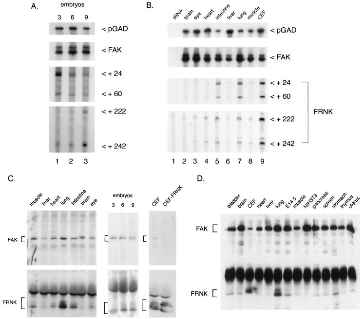
FIG. 3.
FRNK expression during chicken embryogenesis. RNA samples were prepared from either cultured CEF, whole chicken embryos (day 3, 6, or 9), or tissues isolated from day 18 chicken embryos as described in Materials and Methods. Individual RNA samples were hybridized to FAK-, FRNK-, and GAD-specific RNA probes and analyzed as described in the legend to Fig. 1. (A) RNA from day 3 (lane 1), day 6 (lane 2), and day 9 (lane 3) whole chicken embryos. Exposure times for the autoradiograms were 24 h for FAK, 1 week for FRNK, and 4 h for pGAD. (B) RNAs from 18-day-old chicken embryo brain (lane 2), eye (lane 3), heart (lane 4), intestine (lane 5), liver (lane 6), lung (lane 7), and muscle (lane 8). Exposure times were 2 days for FAK and FRNK and 3 h for pGAD. (C) Expression of FAK and FRNK proteins in chicken tissues and whole embryos. One milligram individual tissues or cultured cells and 3 mg (whole embryos) of cell extracts were immunoprecipitated with the FAK/FRNK-specific antibody BC3 as described in Materials and Methods. Detection of FAK and FRNK with BC3 was optimized by separating upper and lower halves of the membrane and immunoblotting each portion of the membrane as described in Materials and Methods. CEF ectopically expressing (CEF-FRNK) were used as positive controls for FRNK expression. The positions of FAK- and FRNK-specific proteins are indicated by brackets. The slower-migrating form of FAK present in brain is likely encoded by an alternatively spliced FAK transcript identified by sequence analysis of brain cDNAs (4). (D) Expression of FAK and FRNK proteins in murine cells and tissues. One-half milligram of lysate protein from each cell and tissue type was immunoprecipitated and immunoblotted with a polyclonal antibody raised against murine FAK sequences, FAK C-20 (see Materials and Methods).

