Figure 1.

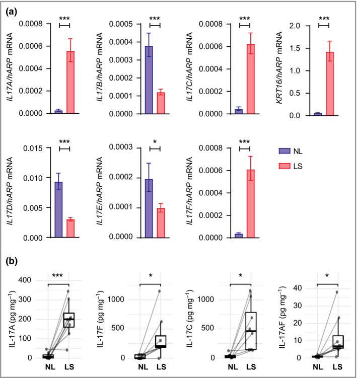
Interleukin (IL)‐17A, IL‐17F, IL‐17A/F and IL‐17C are overexpressed in lesional psoriasis skin. (a) Gene expression of IL17 family cytokines and KRT16 (positive control) in paired samples of lesional and nonlesional skin (n = 10) analysed by quantitative polymerase chain reaction. hARP (RPLP0) was used as the reference gene for normalization. Bars show mean mRNA expression (2ΔCt) (SEM). P‐values were calculated via marginal means (i.e. least square means) and were Benjamini–Hochberg adjusted for multiple testing. (b) Protein levels of IL‐17 family cytokines measured in paired samples of lesional and nonlesional skin (n = 8) using enzyme‐linked immunosorbent assay or MSD kits (Meso Scale Discovery). Protein levels (pg mg–1 total protein) are plotted as means with lower and upper hinges corresponding to the 25th and 75th percentiles. P‐values were calculated using a linear mixed‐effects model. *P < 0·05, **P < 0·01 and ***P < 0·001. LS, lesional skin; NL, nonlesional skin.
