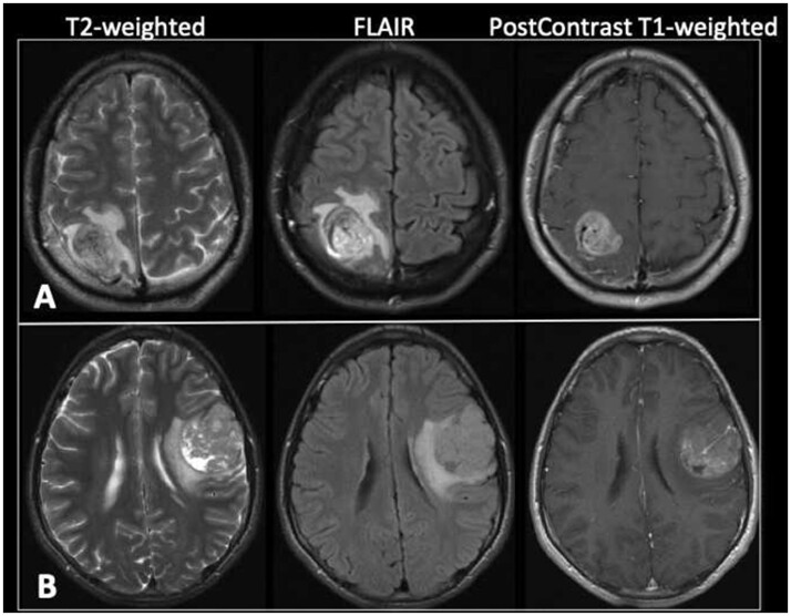
FIGURE 6.
Preoperative MR images of subjects with anaplastic PXA that had tumor progression. (A) Axial T2-weighted, FLAIR, and postcontrast T1-weighted images of subject 3 at the time of initial presentation demonstrate a heterogeneously enhancing right parietal lobe mass with surrounding vasogenic edema and mild local mass effect. A prominent vessel is noted coursing through the mass. (B) Axial T2-weighted, FLAIR, and postcontrast T1-weighted images of subject 12 at the time of initial presentation demonstrate a heterogeneous predominantly enhancing mass in the posterior left frontal lobe. There are a few small cystic areas within the mass. A 5-mm-thick area of increased T2 signal deep to the tumor is consistent with vasogenic edema. There is mild regional mass effect including on the left lateral ventricle. A prominent vessel is seen within the mass.

