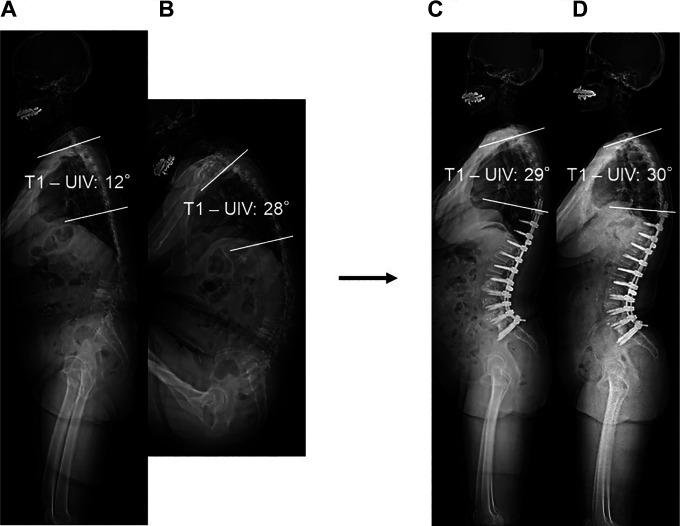
Figure 3.
Time course of T1-UIV after correction surgery in a patient with Ideal group. (a) Baseline preoperative radiograph in standing position. (b) Whole spine full flexion lateral radiograph. (c) Postoperative radiograph. (d) Final follow-up radiograph. These X-ray images demonstrating reciprocal changes and whole spine full flexion lateral radiograph correctly estimate the postoperative alignment of unfused thoracic spine. Abbreviations: T1-UIV, angle between T1 and UI.V.

