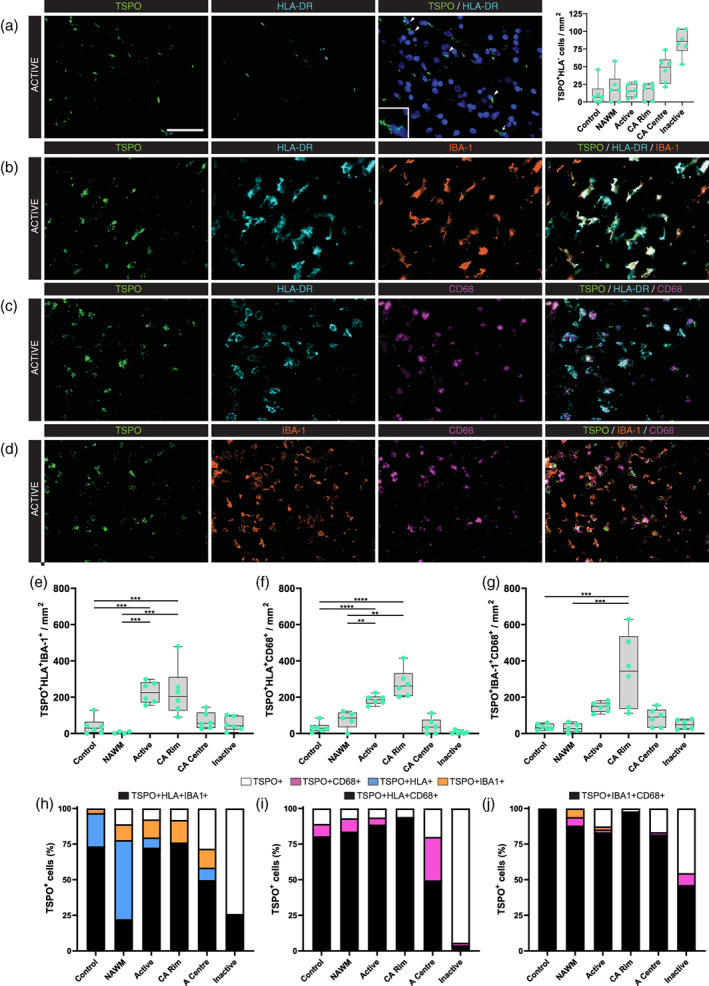
FIGURE 3.

Co‐localization of TSPO with microglia/macrophage markers in white matter lesions in MS. Representative images of TSPO + HLA‐DR− cells in active lesion areas in MS (a), and representative images showing co‐localization between TSPO and HLA‐DR/IBA‐1 (b), HLA‐DR/CD68 (c), IBA‐1/CD68 (d). Quantitative analysis showed an increase in microglia markers in active lesion areas for all different staining combinations (e–g). Percentages showing that nearly all TSPO+ cells in white matter MS lesions co‐localize with at least one or two microglia/macrophage markers (h–j). **p < .01, ***p < .001, ****p < .0001. Scale bar = 50 μm. CA, chronic active; NAWM, normal appearing white matter
