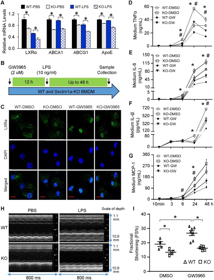
Figure 6.
LXR agonist fails to rescue LPS-induced inflammation and cardiac dysfunction in Sectm1a KO model. (A) Gene expression of LXRα and target genes in WT and Sectm1a-KO BMDMs after 3 h of LPS (10 ng/mL) treatment was measured using qRT-PCR (n = 3). (B) Graphic scheme of treatment protocol. BMDMs from WT and Sectm1a KO mice were first treated with LXR agonist, GW3965 (2 µm, 12 h) followed by LPS stimulation (10 mg/mL, up to 48 h). (C) Immunofluorescent staining of BMDMs with LXRα after 12 h GW3965 and 30 min LPS treatment. DNA was stained with DAPI (blue). (D–G) After treating BMDMs with GW3965 for 12 h, cytokine levels in cell culture supernatant was determined by ELISA at indicated time points post-LPS treatment (n = 4–5). (H and I) WT and Sectm1a KO mice received three injection of GW3965 (30 mg/kg of BW, once daily, DMSO used as control), 6 h after last GW3965 injection, all mice received LPS injection (10 mg/kg) and underwent echocardiography measurement to assess cardiac function (n = 4–7) (Scale bar, 10 µm; *P < 0.05 when comparing WT-DMSO to WT-GW, #P < 0.05 when comparing WT-GW to KO-GW; data are presented as mean ± SEM; two-way ANOVA).

