Figure 7.
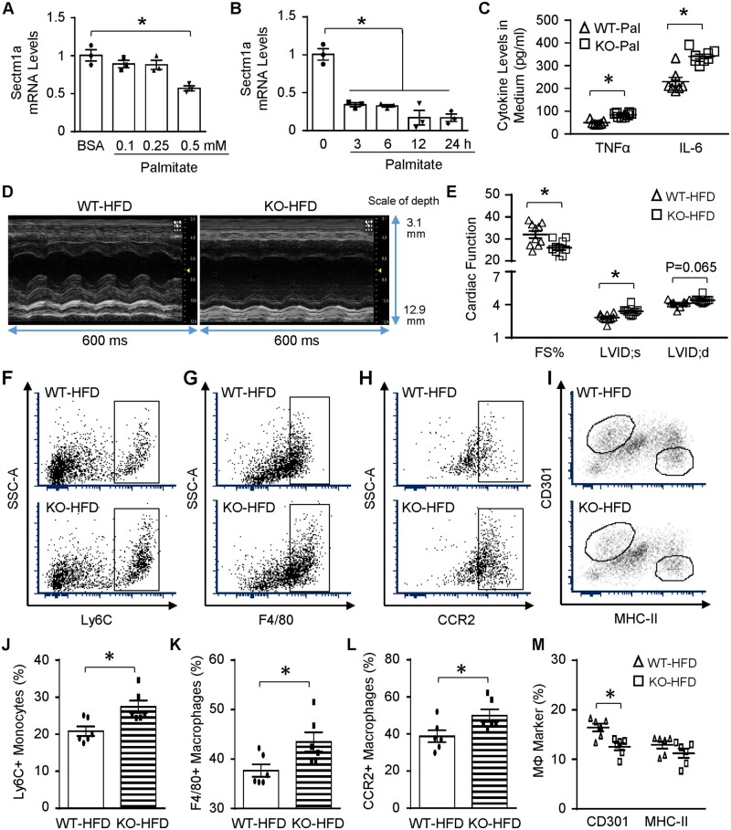
Lack of Sectm1a promotes HFD-induced cardiac inflammation and dysfunction. (A) WT BMDMs were treated with indicated doses of palmitate (A) for 24 h and (B) RAW264.7 macrophages were treated with 0.5 mM palmitate for indicated time points, then gene expression of Sectm1a was measured with qRT-PCR (n = 3). (C) WT and Sectm1a KO BMDMs were treated with palmitate (0.5 mM, 24 h), and cytokine levels in cell culture supernatant were measured using ELISA (n = 7–8). (D and E) Cardiac function was determined by echocardiography after WT and Sectm1a KO mice were fed with HFD for 20 weeks (n = 10 per group). (F–M) Representative flow cytometry plots and quantification of cardiac macrophage marker expression showed more monocytes (Ly6C+) and macrophage accumulation (F4/80+) with inflammatory phenotype (CCR2+, CD301-) in the heart of KO mice 5 weeks after HFD feeding (n = 6). FS, fractional shortening; LVID;d, left ventricular internal diameter at diastole; LVID;s, left ventricular internal diameter at systole (*P < 0.05; data are presented as mean ± SEM; Student’s t-test).

