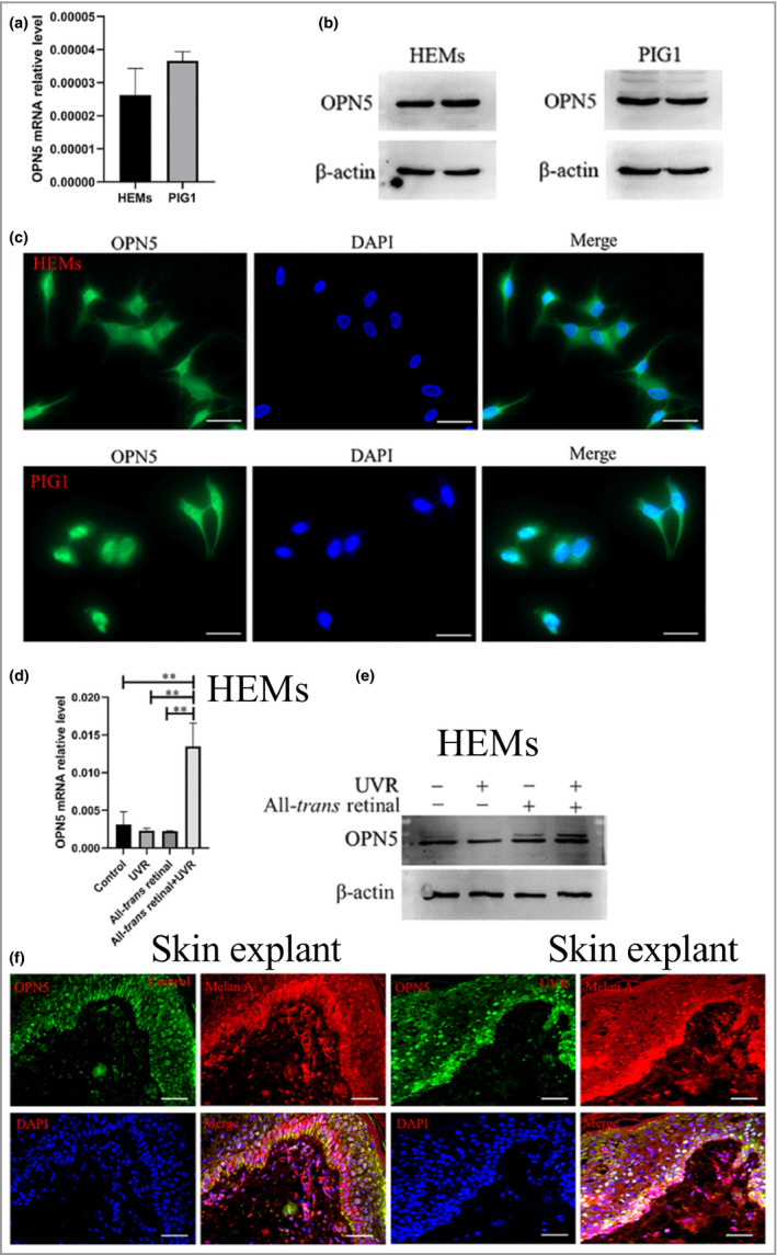
Figure 1.

Opsin 5 (OPN5) senses ultraviolet radiation (UVR) in human epidermal melanocytes (HEMs). (a) OPN5 mRNA levels were normalized to glyceraldehyde 3‐phosphate dehydrogenase (GAPDH) levels. (b) Western blot analysis of OPN5 protein expression in HEMs and transfected human melanocyte line PIG1. (c) Representative images show OPN5 protein expression in HEMs (top) and PIG1 cells (bottom). The left panel represents localization of opsins (Alexa Fluor 488, green) at the plasma membrane, the middle panel represents the nucleus stained with 4′,6‐diamidino‐2‐phenylindole (DAPI) (blue), and the right panel represents an overlay of the middle panel and the phase contrast image. Scale bars = 20 µm. (d, e) HEMs were irradiated with UVR, and expression levels of OPN5 gene and protein were determined by quantitative real‐time reverse‐transcription polymerase chain reaction and Western blot analysis. All data are shown as mean ± SEM of three independent experiments. Beta‐actin was used as loading control. Statistical significance was determined by one‐way anova. **P < 0·01. (f) OPN5 expression (Alexa Fluor 488, green) colocalized with melanocyte marker MelanA (Cy3, red) in skin explant with immunofluorescence staining, without UVR (left) or with UVR (right). Scale bars = 50 µm.
