Figure 2.

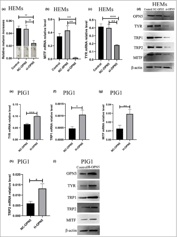
Opsin 5 (OPN5) mediates melanogenesis. (a) Human epidermal melanocytes (HEMs) were transfected with small interfering (si)RNA directed against OPN5 (si‐OPN5), or a negative control (NC‐OPN‐RNA). After short interfering (si)RNA inhibited OPN5, melanin changes in HEMs were measured by the NaOH method. (b, c) After siRNA inhibited OPN5, quantitative real‐time reverse‐transcription quantitative polymerase chain reaction (RT‐qPCR) was used to analyse changes of tyrosinase (TYR) and microphthalmia‐associated transcription factor (MITF) gene expression in HEMs. (d) After siRNA inhibited OPN5, Western blotting was used to analyse the changes of TYR, tyrosinase‐related protein 1 (TRP1), TRP2 and MITF protein expression in HEMs. Beta‐actin was used as a loading control. (e–i) After OPN5 was overexpressed (as high‐expression H‐OPN5‐RNAi) in PIG1 cells, RT‐qPCR and Western blotting were used to analyse gene and protein expression levels of TYR, TRP1, TRP2 and MITF. Beta‐actin was used as loading control. All data are shown as mean ± SEM of three independent experiments. Statistical significance was determined by one‐way anova followed by Tukey’s test. *P < 0·05, **P < 0·01, ***P < 0·001.
