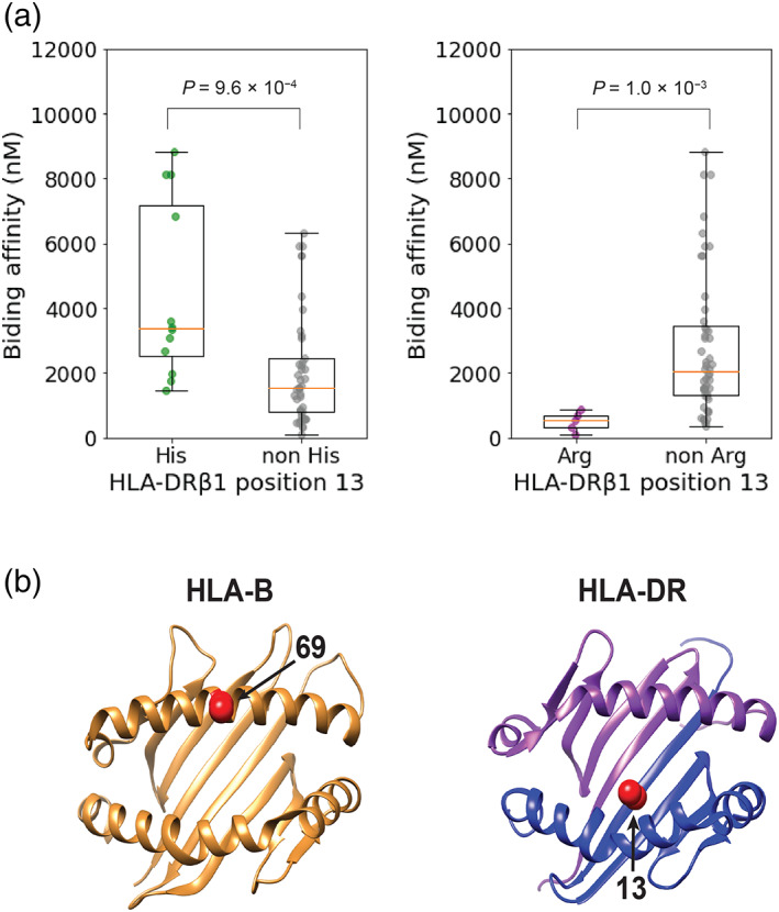
FIG. 3.

Risk‐associated amino acid polymorphisms and in silico predicted binding affinity of HLA‐DRB1 alleles to an α‐synuclein epitope. (a) The results of in silico predicted binding affinity of HLA‐DRB1 alleles to the Y39 epitope of α‐synuclein are shown in boxplots and compared between alleles with and without His (left) and Arg (right) in position 13 of HLA‐DRβ1. His13 in HLA‐DRβ1 is almost consistent with HLA‐DRB1*04, and HLA‐DRB1*15:01 has Arg13 in HLA‐DRβ1. (b) Three‐dimensional ribbon models of the human leukocyte antigen (HLA) proteins associated with Parkinson's disease (PD) risk. The protein structures of HLA‐B and HLA‐DR are based on Protein Data Bank entries 2BVP and 3PDO, respectively, which were displayed using UCSF Chimera version 1.14. Residues at the PD risk‐associated amino acid positions are colored red (arrows). Arg, arginine; His, histidine. [Color figure can be viewed at wileyonlinelibrary.com]
