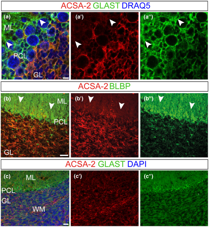
FIGURE 1.

ACSA‐2 presents a unique expression pattern in the adult cerebellum. Coronal (a) and sagittal (b,c) adult mouse (younger than 2 months) cerebellar sections were used to investigate ACSA‐2 expression. Confocal stacks identified ACSA‐2, unlike the common astrocyte markers GLAST (a,c) and BLBP (b), to be distinct in the three cerebellar cortical layers (a,c). GLAST+ BLBP+ BG processes in the ML (a″,b″,c″; arrowheads in a″,b″) revealed weak ACSA‐2 expression (a′–c′; arrowheads in a′,b′). High ACSA‐2 expression was found on velate protoplasmic astrocytes in the GL (a′–c′). ACSA‐2 is further expressed by a third type of cerebellar glia, the fibrous astrocytes in the WM (c). Nuclear stain: DRAQ5 (a), DAPI (c). Scale bars: 5 μm (a), 50 μm (b) 30 μm (c). GL, granular cell layer; ML, molecular layer; PCL, Purkinje cell layer; WM, white matter
