FIGURE 2.

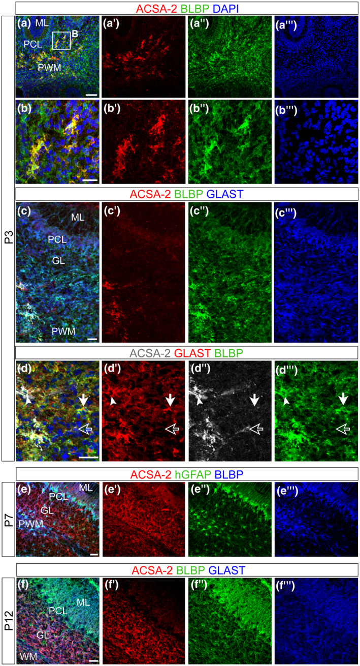
ACSA‐2 defines a subpopulation of astrocyte progenitors in the neonatal cerebellum. The ontogeny of ACSA‐2+ cells was investigated using confocal stacks of P3 (a–d), P7 (e) and P12 (f) sagittal sections. At P3, ACSA‐2 was not detected in the nascent ML (a′,c′) and neither in the PCL (a′,c′). BG progenitors, which are marked by BLBP and GLAST (a″,c″,c″′), are not expressing ACSA‐2. ACSA‐2 is present in the PWM (a′,b′,c′,d″) but it is not as broadly expressed as the common markers GLAST and BLBP (a″,c″,c″′,d′,d″′). Within the GLAST+ population (of the PWM) ACSA‐2 positive (white arrowheads in d) as well as ACSA‐2 negative cells (filled white arrows in d) and a very rare population of ACSA‐2+/GLAST− cells (empty arrows in d) were detected (d–d″). At P7, the labeling of ACSA‐2 appeared in the PWM and the GL and overlapped with the reporter expression of the human glial fibrillary acidic protein (hGFAP) in the PWM and the GL (e). At P12, ACSA‐2 expression has expanded to include BLBP+ and GLAST+ cells in both inner compartments: the GL and the WM (f). DAPI, nuclear stain. Scale bars: 100 μm (a), 30 μm (c,e,f), 50 μm (b,d). GL, granular cell layer; ML, molecular layer; PCL, Purkinje cell layer; PWM, prospective white matter; WM, white matter
