
FIGURE 7.
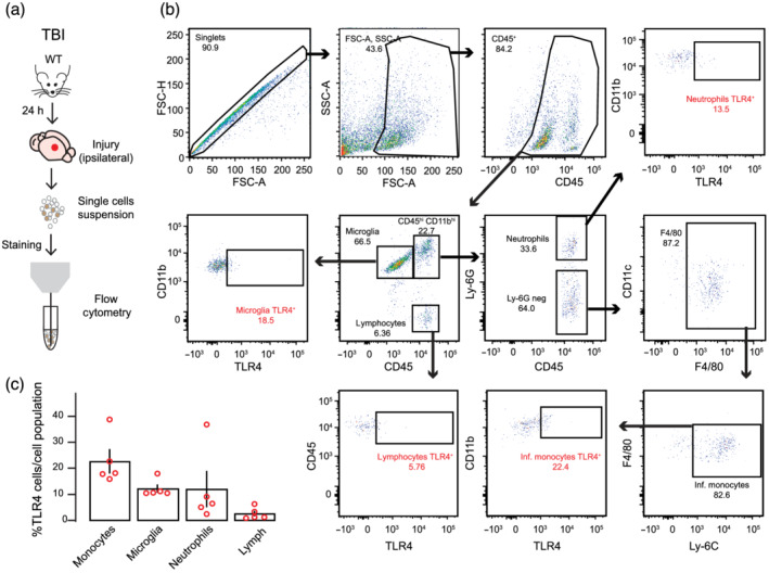
Infiltrating monocytes and resident microglia exhibits TLR4 receptors in injured cortex. (a) Schematic of the experimental protocol. (b) Flow cytometry analysis of immune cells gated on CD45+ from a representative TBI ipsilateral cortex. (c) Quantification of the percentage of cell types expressing TLR4 receptors of each cell population among CD45+ cells (microglia: CD11bintCD45int; lymphocytes: CD11bnegCD45hi; neutrophils: CD11bhiCD45hiLy‐6Ghi; and infiltrating monocytes: CD11bhiCD45hiLy‐6G‐F4/80 + Ly‐6Chi). Open dots on scatterplots are representative of each ipsilateral brain of n = 5 mice and depict the % of TLR4+ cells exhibiting extracellular receptor expression within each population