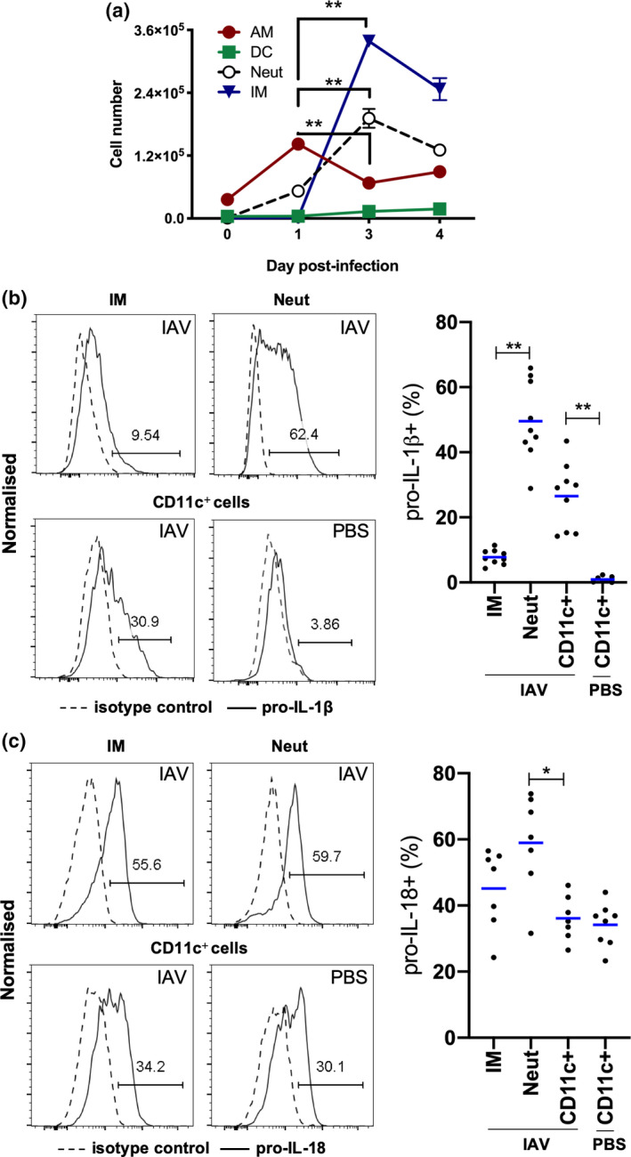
Figure 3.

Expression of pro‐IL‐1β and pro‐IL‐18 by innate immune cells in BAL. (a–c) C57BL/6 mice were infected with 105 PFU of HKx31 and BAL cells were isolated and characterized by flow cytometry. (a) Numbers of CD11c+I‐Ablow AMs, CD11c+I‐Abhigh DCs, Ly6G+ neutrophils (Neut) and Ly6C+ IMs on days 0, 1, 3 and 4 postinfection. Mean ± s.e.m. **P < 0.01; day 1 versus day 3 only shown. (b) Representative histograms showing the expression of pro‐IL‐1β by BAL IMs, Neut and CD11c+ cells from uninfected PBS‐treated (PBS) or IAV‐infected mice on day 4. Proportion of pro‐IL‐1β+ IMs, Neut and CD11c+ cells in BAL. (c) Representative histograms showing expression of pro‐IL‐18 by BAL IMs, Neut and CD11c+ cells from uninfected PBS‐treated (PBS) or IAV‐infected mice on day 4. Proportion of pro‐IL‐18+ IMs, Neut and CD11c+ cells in BAL. Data are relative to isotype control. Individual mice are shown as circles and bars represent the mean. Neut and IMs were not analyzed in uninfected PBS‐controls because of low cell numbers. *P < 0.05, **P < 0.01; one‐way ANOVA. Data are from one of two experiments (n = 7 or 9). AM, alveolar macrophage; BAL, bronchoalveolar lavage; DCs, dendritic cells; IAV, influenza A virus; IL, interleukin; IM, inflammatory macrophage; mRNA, messenger RNA; PBS, phosphate‐buffered saline; PFU, plaque‐forming units.
