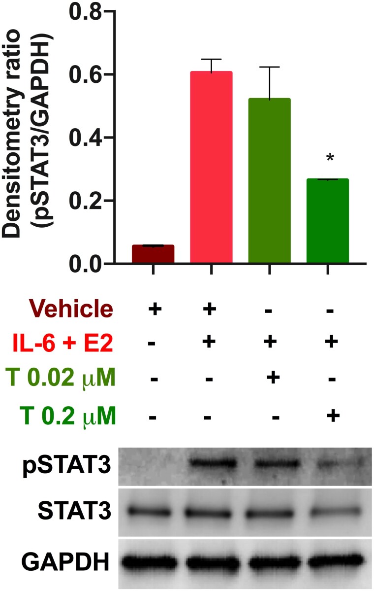
Figure 3.
Tofacitinib reduced IL-6+E2-stimulated phosphorylation of STAT3 in Ishikawa cells. Representative western blot for pSTAT3 levels, GAPDH, and total STAT3 following 24 -h treatment (see Supplementary Information For full western blot image). The ratio of pSTAT3 to GAPDH is compared in the bar graph for exposure with vehicle (dimethylsulphoxide: DMSO), stimulation with IL-6 and estradiol (E2), and treatment with 0.02 or 0.2 µM Tofacitinib. Results shown as mean ± SEM. *P < 0.05 versus stimulation only.

