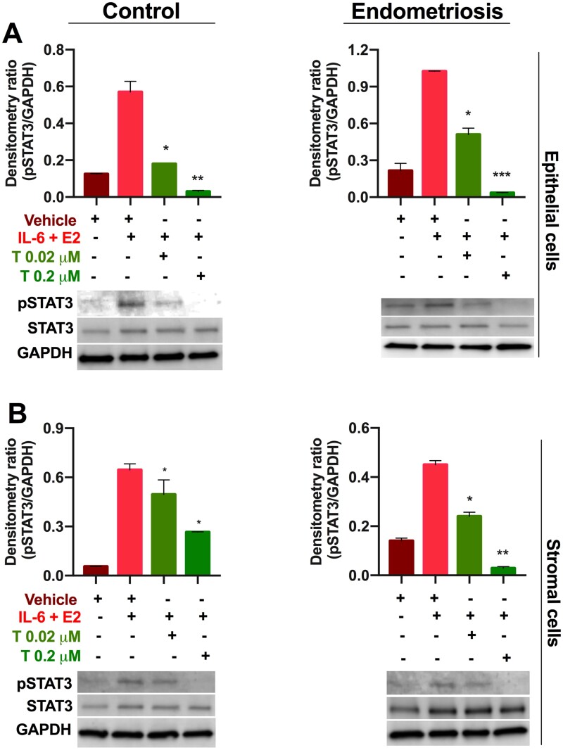
Figure 5.
Tofacitinib treatment decreased pSTAT3 levels in primary human cells. (A) Epithelial cells and (B) stromal cells from patients with (endometriosis) and without (control) endometriosis show that pSTAT levels are increased by IL-6 and E2 treatment while Tofacitinib (0.02 and 0.2 µM) treatment significantly decreased the pSTAT levels stimulated by IL-6 + E2 compared to vehicle (DMSO). Representative western blot images are shown for pSTAT3, STAT3, and GAPDH detection (see Supplementary Information for full western blot images). Bar graph shows the ratio of pSTAT3 to GAPDH by densitometry analysis. Results shown as mean ± SEM. *P < 0.05; **<0.01, and ***<0.001 versus IL-6 + E2 stimulation.

