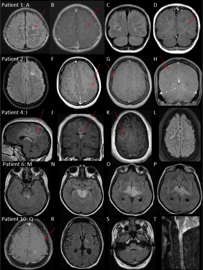
Figure 1: MRI features of dual-positive MOG-IgG1/NMDA-R-IgG patients.

Patient 1, male 2 years.
MRI brain: Axial T2/FLAIR (A) T1 Gd+ (B), coronal T2/FLAIR (C) T1 Gd+ (D) at first attack demonstrating bilateral frontoparietal sulcal T2/FLAIR hyperintensity, diffuse leptomeningeal enhancement and patchy cortical T2/FLAIR hyperintensity.
Patient 2, female 13 years.
MRI brain: Axial T2/FLAIR (E) and T1 Gd+ (F) at third attack age 13, demonstrating focal T2/FLAIR hyperintensity deep to left superior frontal sulcus and in left frontal opercular cortex with left unilateral leptomeningeal enhancement and focus of parenchymal enhancement. Axial T1 Gd+ (G) coronal T1 Gd+(H) at fourth attack age 18, demonstrating right unilateral hemisphere leptomeningeal enhancement and resolution of left frontoparietal leptomeningeal and parenchymal signal abnormalities.
Patient 4, female 14 years
MRI brain: Sagittal T1 Gd+ (I), Coronal T1 Gd+ (J) and Axial T1 Gd+ (K) and Axial T2/FLAIR at first attack demonstrating bilateral (right>left) leptomeningeal enhancement around the splenium corpus and callosum.
Patient 6, female 18 years, Axial T2/FLAIR (M), (N), (O), (P) at second attack 4 weeks from onset. T2/FLAIR hyperintensity of internal capsule, bilateral thalami, midbrain and pons.
Patient 10, male 33 years.
MRI brain: Axial T1 Gd+ (Q) at first attack demonstrating left unilateral leptomeningeal enhancement. Axial T2/FLAIR (R), (S) at second attack 8 weeks later demonstrating T2/FLAIR hyperintensity of the left basal ganglia and bilateral cerebellar peduncles
MRI cervical spine: Sagittal T2 STIR (T), demonstrating cervical myelitis with T2 hyperintensity and cord enlargement from medulla to C4 and associated patchy enhancement (not shown).
