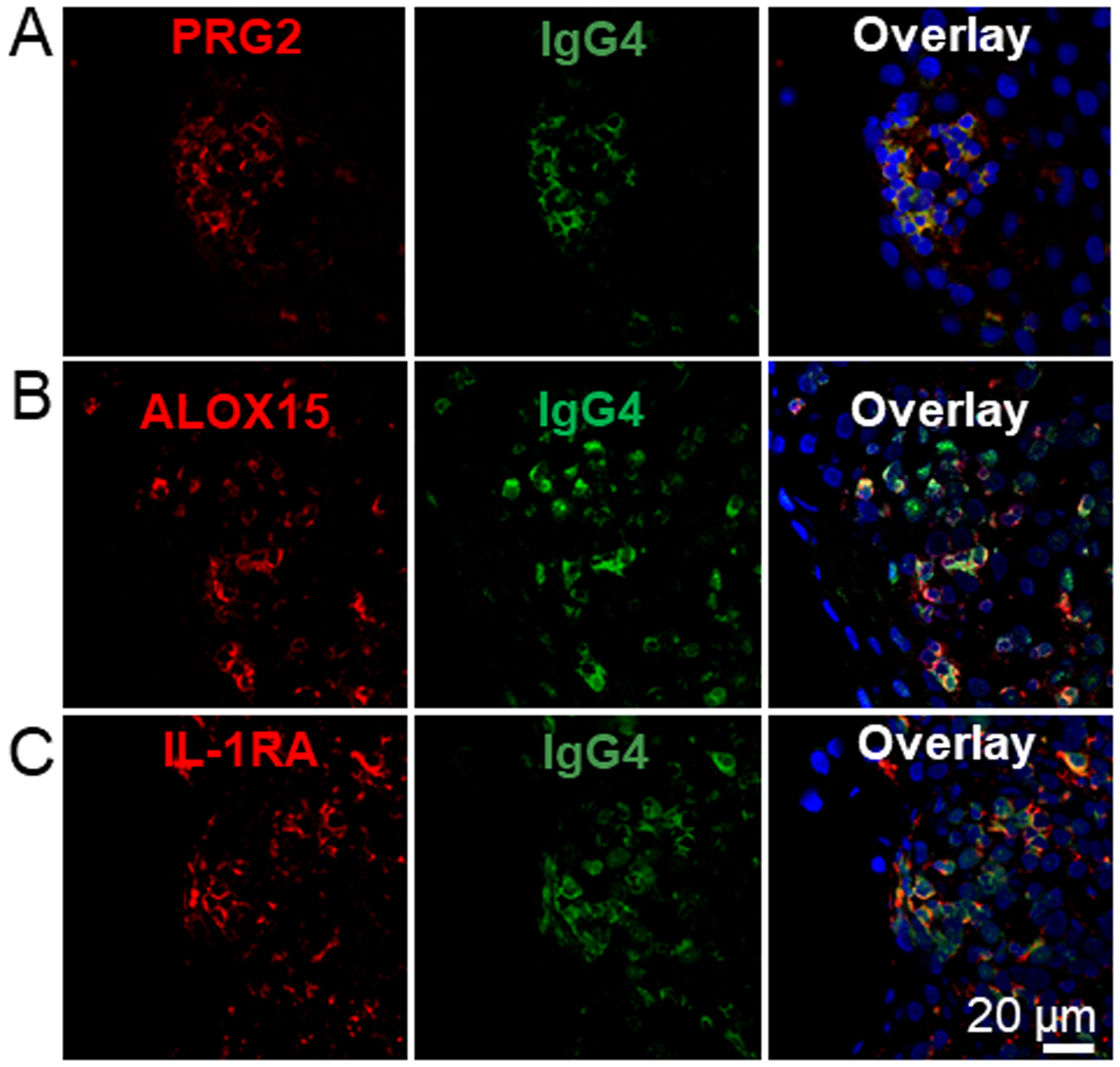
Figure 6. IgG4+ EoE lesions partially co-localize with inflammatory markers.

EoE patient biopsy sections were stained for IgG4 (green) as described in Figure 5 and antibodies specific to the proteins PRG2 (A, red), ALOX15 (B, red) and IL-1RA (C, red). Samples were counter stained with DAPI. N=3.
