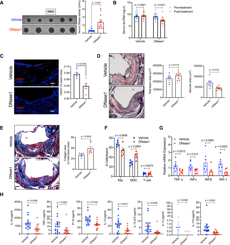
Figure 7.
Exogenous DNase1 administration promotes atherosclerotic plaque remodeling and stabilization. Female Apoe−/− mice were fed western-type diet for 16 wk followed by administration of either vehicle or DNase1 (400 U) intravenously 3× a week for 4 wk. A, Single radial enzyme diffusion analysis of serum total DNase activity in vehicle or DNase1-injected mice. B, Analysis of serum extracellular ds-DNA concentration in indicated groups of mice before and after treatment with vehicle or DNase1. C, Aortic root sections of vehicle and DNase1-treated mice were immunostained with antibody against CitH3 (red) and percent lesion area that stains positive for CitH3 was quantified. Nucleus were stained with DAPI (blue). Bar, 25 μm. n=5 mice per group. D, H&E staining of aortic root sections of vehicle and DNase1-treated mice to quantify the total lesion area and the total necrotic area (red dotted line). Bar, 25 μm. E, Mason trichrome staining of aortic root sections of vehicle and DNase1-treated mice to quantify collagen deposition in the plaque. Bar, 25 μm. F, Immunofluorescence-based quantification of percent macrophages (F4/80), smooth muscle cells (sm-Actin), and T-cells (CD3) in atherosclerotic lesions of vehicle and DNase1-treated mice. G, qPCR-based analysis of relative mRNA expression of indicated proinflammatory genes in aorta of vehicle and DNase1-treated mice. H, Multiplex ELISA for quantification of proinflammatory cytokine levels in the sera of vehicle and DNase1-treated mice. Shapiro-Wilk test was conducted to determine normality of data sets. P were calculated using Mann-Whitney U test (A, C, and E), Wilcoxon paired test (B), or Student t test (D, F–H).

