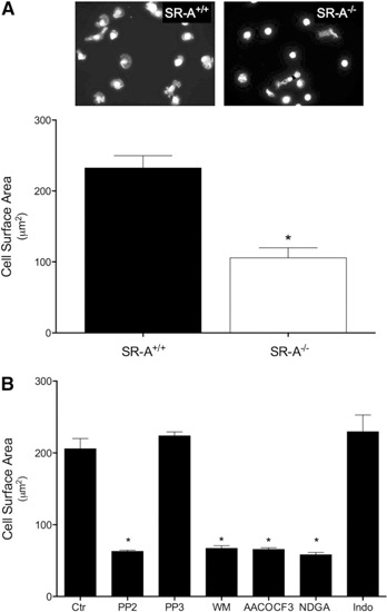
Figure 1.

SR‐A‐mediated macrophage adhesion to gluc‐collagen requires activation of Src kinase, PI3K, and PLA2. (A) Resident MPMs isolated from SR‐A+/+ or SR‐A−/− mice were plated on gluc‐collagen‐coated slides. Cells were adhered for 2 h at 37°C, fixed, and stained with Alexa Fluor568‐conjugated phalloidin. Nuclei were stained with DAPI. Images were digitally captured, and the surface area of individual cells quantified. (B) MPMs isolated from SR‐A+/+ were pretreated as indicated with PP2 (50 µM), PP3 (50 µM), WM (200 nM), AACOCF3 (30 µM), NDGA (50 µM), or indomethacin (Indo; 10 µM) for 30 min in suspension and then plated on gluc‐collagen‐coated slides. Ctr, Control. Cells were stained and cell surface area quantified as described for A. The graphs depict the mean ± sem cell surface area determined in 3 separate experiments. Significant difference (*P < 0.05) from untreated SR‐A+/+ (control) cells.
