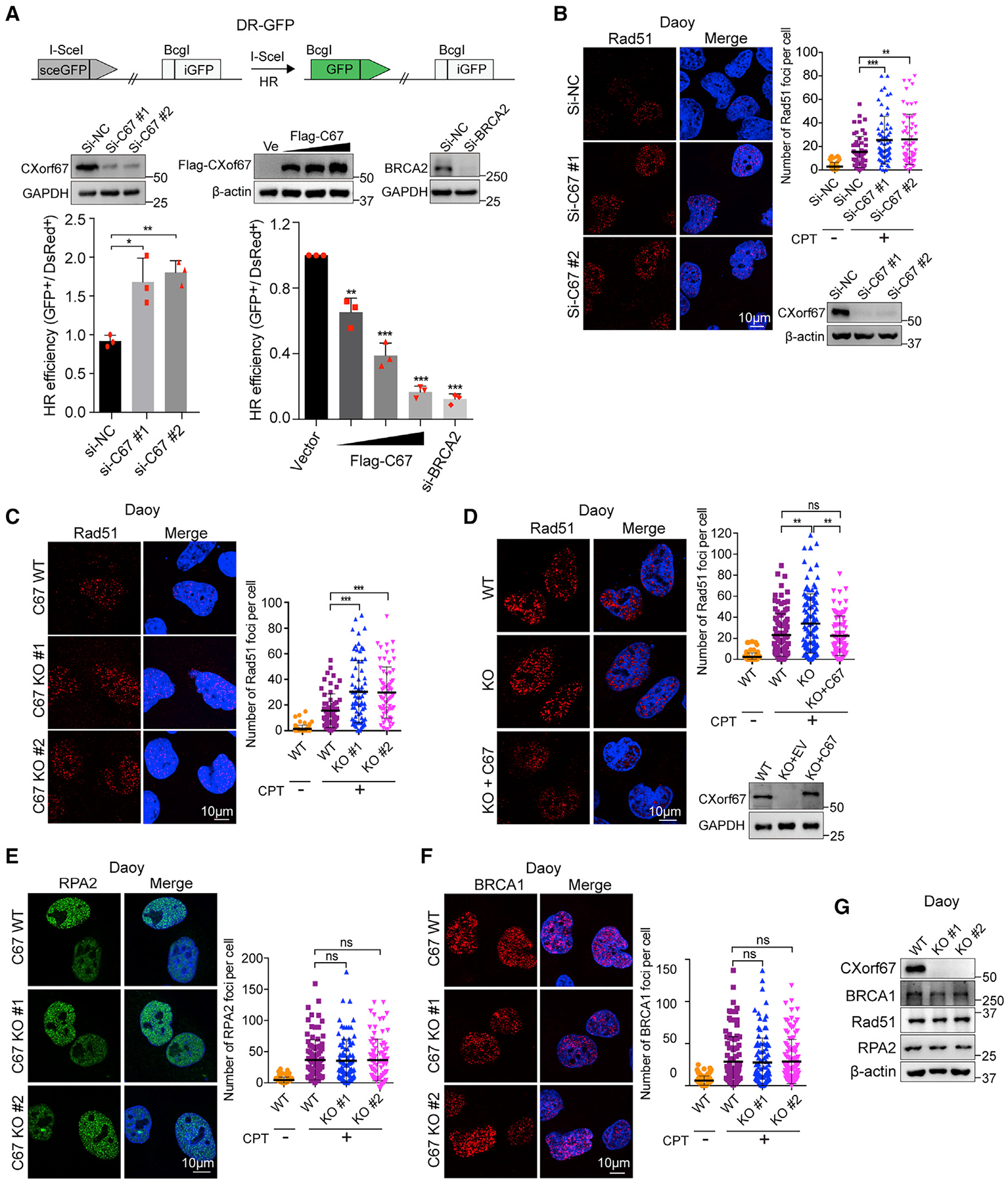
Figure 2. CXorf67 Regulates HR- but Not NHEJ-Mediated DSB Repair.

(A) Schematic illustration of the GFP-based HR reporter assay (DR-GFP). Effect of CXorf67 knockdown and overexpression on the efficiency of homologous recombination (HR) repair. U2OS DR-GFP cells were transfected with CXorf67-specific siRNA or Flag-tagged CXorf67 and control vector together with I-SceI plasmids, and the percentage of GFP-positive cells was analyzed by fluorescence-activated cell sorting 48 h after transfection. Knockdown of BRCA2 act as a positive control for inhibiting HR repair. Data are presented as mean ± SD (n = 3 independent experiments; paired t test; *p < 0.05, **p < 0.01, ***p < 0.001).
(B and C) Knockdown or knockout of CXorf67 leads to increased Rad51 foci formation. Daoy cells were transfected with CXorf67 siRNA or control siRNA for 48 h and then treated with CPT (0.1 μM) for 4 h and immunostained with an RAD51 antibody followed by Cy3-conjugated secondary antibody. Scatter dot plot represents the Rad51 foci per nuclei (right). Data are presented as mean ± SD (n = 56–64; unpaired t test; **p < 0.01, ***p < 0.001). (C) Same as above, but performed in the C67-WT and C67-KO Daoy cells. Scatter dot plot represents the Rad51 foci per nuclei (right). Data are presented as mean ± SD (n = 62–71; unpaired t test; ***p < 0.001).
(D) C67-KO 1# Daoy cells showed increased Rad51 foci formation, which is rescued by re-expression of CXorf67. Cells were treated with CPT (0.1 μM) for 4 h and immunostained with an RAD51 antibody followed by Cy3-conjugated secondary antibody. Scatter dot plot represents the Rad51 foci per nuclei (right). Data are presented as mean ± SD (n = 83–106; unpaired t test; **p < 0.01).
(E and F) Representative images of RPA2 foci and BRCA1 foci in Daoy C67-WT and C67-KO cells that were treated with CPT (0.1 μM) for 4 h. Quantification of RPA2 and BRCA1 foci per nuclei using the Image-Pro Plus software. Scatter dot plot represents the RPA2 foci per nuclei. Data are presented as mean ± SD (n = 88–96; unpaired t test; ns, not significant, p > 0.05) (E). Data are presented as mean ± SD (n = 98–110; unpaired t test; ns, not significant) (F).
(G) CXorf67, Rad51, RPA2, and BRCA1 gene expressions at the protein level in C67-WT and C67-KO Daoy cells were validated by western blotting.
See also Figure S2.
