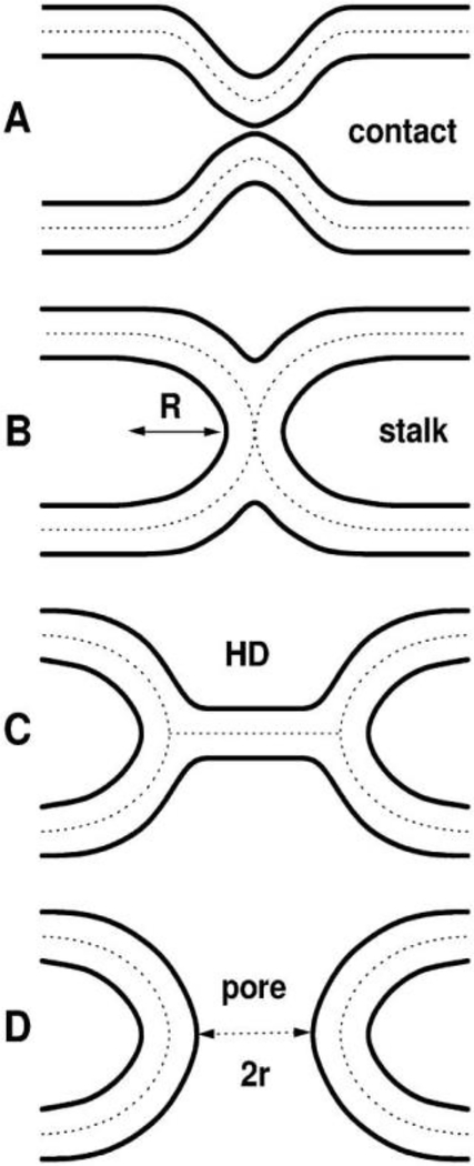
Figure 6.
Well-known hypothetical fusion intermediates. Lipid bilayer surfaces are indicated by solid lines. Dotted lines in the hydrocarbon interior divide the bilayers into monolayers. A. Contact of the virus and target membranes, B. stalk that allows lipid mixing, C. hemifusion diaphragm (HD), D. pore. Figure from Ref. [2].

