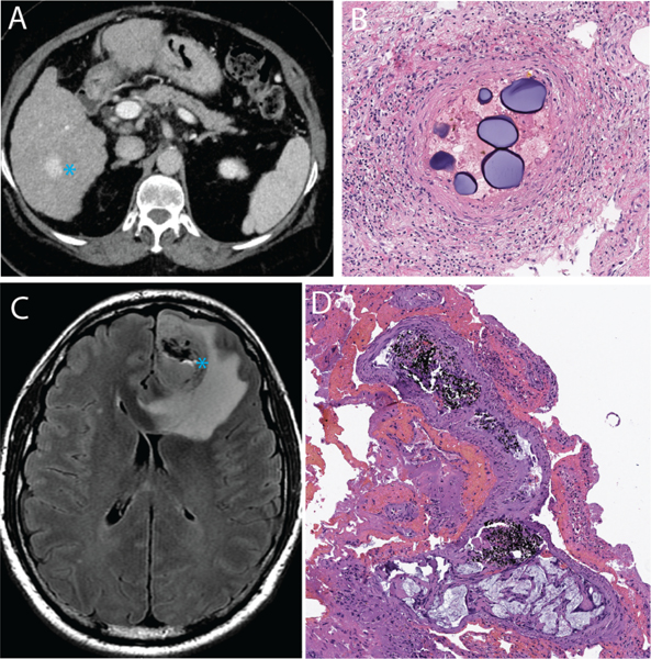
Figure 1.
Clinical examples of embolic agents. A, a patient with cirrhosis and hepatocellular carcinoma (blue asterisk) was treated with microsphere embolization. B, Post-surgical histologic evaluation of the tumor revealed intravascular localization of the microspheres with adjacent inflammatory reaction. C, a patient with glioblastoma multiforme underwent pre-surgical embolization with a liquid embolic agent (Onyx). D, the black-colored liquid embolic agent could be seen within the tumor on the post-surgical resection specimen.

