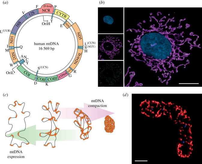
Figure 1.
mtDNA structure, distribution and packaging. (a) Map of human mitochondrial DNA. The loci of genes encoded on the L-strand (inner circle) and the H-strand (outer circle) are indicated. (b) Distribution of mtDNA nucleoids within a human cell. Representative super-resolution Airyscan image of a HeLa cell, with the mitochondrial network labelled using an antibody against the outer membrane protein TOM20 (magenta), mtDNA nucleoids labelled with an anti-DNA antibody (green) and the nucleus stained using DAPI (blue). Merged and single channels are shown. Scale bar represents 10 µm. (c) Packaging of mtDNA by TFAM. The binding of TFAM bends and compacts mtDNA to form the nucleoid. Greater concentrations of TFAM create increasingly compacted nucleoids that are unable to undergo transcription and replication. (d) Three-dimensional rendered super-resolution microscopy image of packaged mtDNA nucleoids within a mitochondrion. A three-dimensional cross-section of TOM20 (red) and mtDNA nucleoids (blue) was acquired using STED microscopy in a HeLa cell. To visualize the nucleoids as three-dimensional objects, the acquired z-stack was deconvolved and rendered using the surface render function within the Huygens Essential software package. Scale bar represents 0.5 µm.

