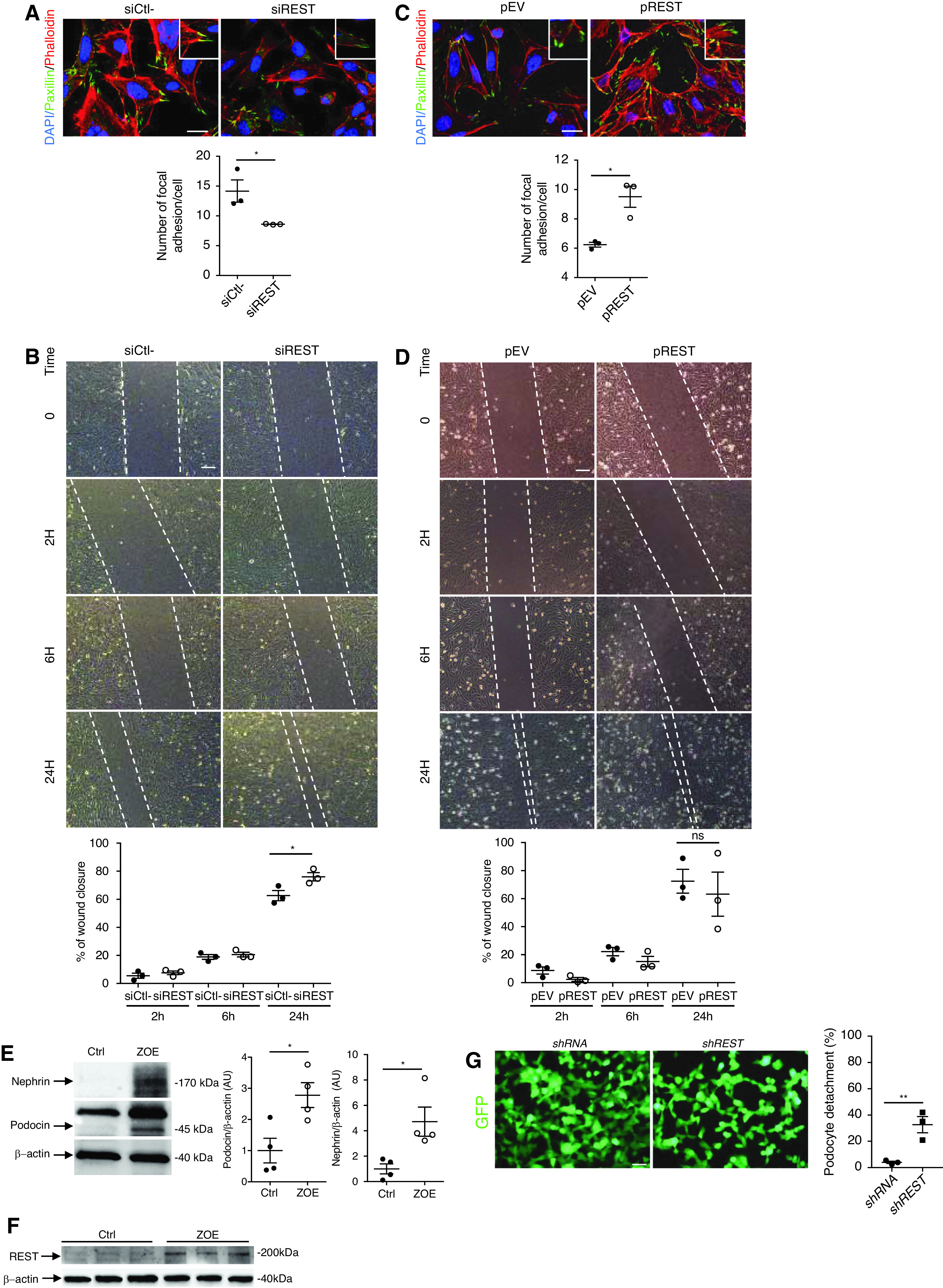
Figure 4.

REST regulates podocyte migration and attachment to the basement membrane. (A) Coimmunostaining using phalloidin, an F-actin marker, and paxillin-specific antibodies and quantification in immortalized mouse podocyte transfected with siREST or siCtrl (n=3 experiments). Scale bars, 10 μm. The insets show higher magnification images of podocyte structures with stress fibers and focal adhesions. (B) Wound healing assay (upper left) and quantification of percentage of wound closure after 2 hours, 6 hours, and 24 hours (lower left) in immortalized mouse podocytes transfected with either siREST or siCtrl (n=3 experiments). Scale bars, 5 μm. (C) Coimmunostaining using phalloidin, an F-actin marker, and paxillin-specific antibodies in immortalized mouse podocytes transfected with either pREST or pEV (n=3 experiments). Scale bars, 10 μm. The insets show higher magnification images of podocyte structures with stress fibers and focal adhesions in pREST-transfected mouse podocytes. (D) Wound healing assay (upper right) and quantification of percentage of wound closure (lower right) after 2 hours, 6 hours, and 24 hours using immortalized mouse podocytes transfected with either pREST or pEV (n=3 experiments). Scale bars, 5 μm. Western blot and quantification of nephrin (E), podocin (E), and REST (F) in immortalized mouse podocytes exposed to mechanical stress and flow over 8 days (ZOE organ-on-a-chip) or maintained under normal static culture conditions (Ctrl). (G) Immortalized podocytes transfected with either scrambled shRNA or REST shRNA. Both shRNAs coexpress GFP. Cells transfected with REST shRNA show greater detachment under mechanical and flow constraints compared with controls (n=3). Quantification shows the percentage of detached cells before and after mechanical/flow stress. Scale bar, 5 μm. All data are shown as the mean±SEM. *P<0.05, **P<0.01, ***P<0.001, ***P<0.0001 between groups, Mann–Whitney, or t test.
