Figure 6.

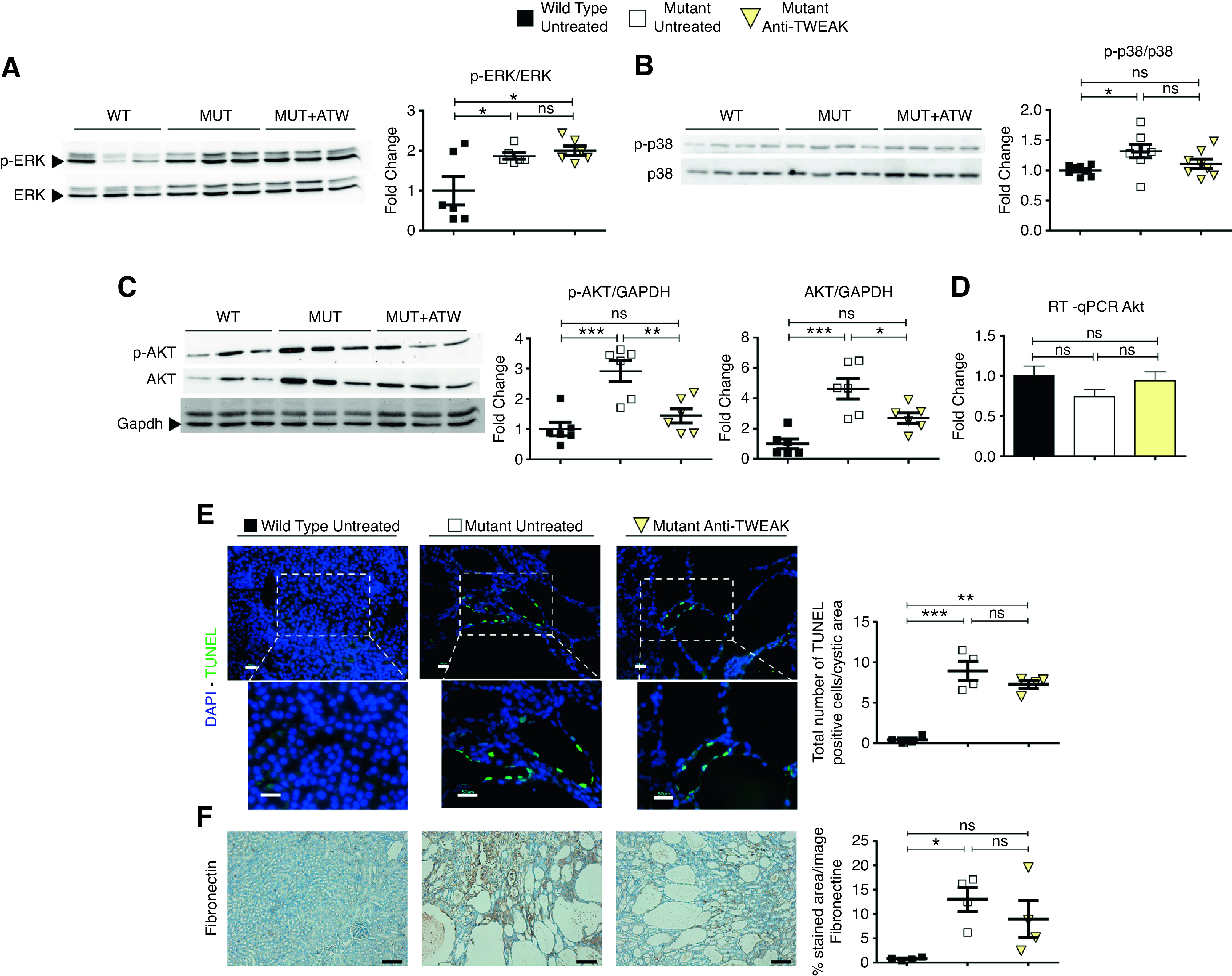
AntiTWEAK decreases cell proliferation via inhibition of AKT pathway. Anti-TWEAK treatment reduces AKT and p-AKT, but not p-ERK, at p30. (A) Western blot analysis for p-ERK and ERK of total kidney protein lysates of WT (n=6), mutant (n=6), and mutant anti-TWEAK–treated mice (n=6). (Right) p-ERK/ERK ratio. (B) Western Blot analysis for p-p38 and p38 of total kidney protein lysates of WT (n=6), mutant (n=6), and mutant anti-TWEAK–treated mice (n=6). (Right) p-p38/p38 ratio. (C) Western blot for p-AKT, AKT, and GAPDH of total kidney protein lysates of WT (n=6), mutant (n=6), and mutant anti-TWEAK–treated mice (n=6). (Right) Ratios for p-AKT/GAPDH, and AKT/GAPDH. GAPDH was used as a housekeeping gene because of changes in AKT protein levels, which precluded the use of AKT to normalize p-AKT levels. (D) RT-qPCR of Akt1 gene for WT (n=5), mutant (n=5), and mutant animals treated with anti-TWEAK (n=6). No significant differences in Akt1 mRNA were observed. (E) Anti-TWEAK decreases apoptosis in kidney anti-TWEAK–treated group, with weak significance (P<0.1), in the cystic area, as detected by TUNEL assay. Scale bars, 100 µm and 50 µm. Original magnification, ×200 and ×400. (F) Immunohistochemical analysis of fibronectin (center) in kidney slides of WT, mutant, and anti-TWEAK groups. n=4 was used per group. Fibrosis, assayed by fibronectin staining, was modestly diminished by anti-TWEAK treatment. Scale bars, 100 µm. Mice were euthanized at p30, according of the scheme shown in Supplemental Figure 4C (medium-term model). Bars represent means±SEMs. *P<0.05, **P<0.01, ***P<0.001. DAPI, 4′,6-diamidino-2-phenylindole.
